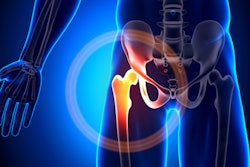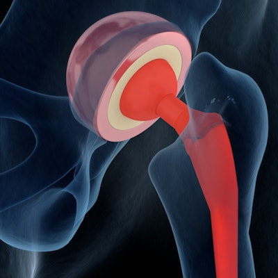
Dual-energy CT performs better than x-ray when it comes to diagnosing loosened hip prostheses, according to a study published July 6 in Radiology.
The findings suggest that clinicians have another effective tool in their arsenal for identifying unstable hip prostheses, wrote a team led by Dr. Giovanni Foti of IRCCS Sacro Cuore Don Calabria Hospital in Negrar, Italy.
"Revisions of hip prostheses are increasing, and conventional radiography is a primary tool for managing complications," the group wrote. "However, dual-energy CT with virtual monoenergetic imaging is capable of reducing periprosthetic metal artifacts compared with standard CT."
Foti and colleagues conducted a study that included 178 patients treated for painful hip prostheses between January 2018 and October 2020. All underwent x-ray and dual-energy CT exams; 49% were diagnosed with loosened prostheses. Two readers blinded to clinical findings evaluated the images and the researchers calculated sensitivity and specificity for each modality.
Dual-energy CT had higher sensitivity than x-ray for both readers (94% and 92%, compared with 84% and 80%). Specificity was comparable, at 93% and 95% for DECT compared with 91% for both for x-ray.
"Dual-energy CT showed better diagnostic performance than conventional radiography in diagnosing hip prosthesis loosening," the researchers concluded.