When it comes to imaging diseases like COVID-19, FDG-PET/CT may not be the first modality that springs to mind. But its performance over the past year for identifying infection and inflammation in COVID-19 patients offers strong evidence of its value.
A group of U.S. nuclear medicine specialists and a colleague in Iran assessed the use of FDG-PET/CT in COVID-19 studies last year and found ample evidence suggesting its effectiveness for diagnosing the disease, clarifying extrapulmonary manifestations, and tracking treatment response. The team shared their findings in a literature review published June 22 in Seminars in Nuclear Medicine.
"While standard radiologic imaging techniques, mainly chest x-ray (CXR) and CT, are the primary imaging modalities to diagnose lung infection, FDG-PET/CT has proven to be effective in cases where conventional imaging falls short," wrote lead author Dr. Ali Gholamrezanezhad of the University of Southern California in Los Angeles.
In their review, Gholamrezanezhad and colleagues outlined FDG-PET/CT's value for elucidating COVID-19 lung infections.
Detecting undiagnosed cases
While reverse transcription polymerase chain reaction (RT-PCR) tests, chest x-ray, and chest CT have been the primary tools of diagnosis, FDG-PET/CT detected undiagnosed cases of COVID-19 in several studies the early stages of infection, when symptoms were nonspecific.
One series of seven patients with two consecutive negative RT-PCR tests found that pulmonary lesions in COVID-19 patients displayed significantly increased SUVmax values when compared with the lungs of control subjects (3.44 versus 0.54). Another study showed that FDG-PET/CT imaged new lung infiltrates in asymptomatic patients approximately seven days prior to the onset of symptoms.
"Routine staging FDG-PET/CT scans may show incidental but typical imaging findings of COVID -19 and help initiate further workup," Gholamrezanezhad wrote.
Pneumonia
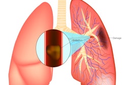
COVID-19 pneumonia is FDG-avid and numerous studies documented lung uptake in both asymptomatic and symptomatic patients.
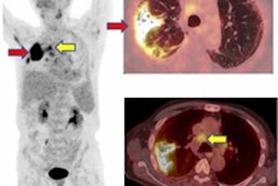
One of the earliest case reports of PET/CT imaging of COVID-19 described four presumed cases with pulmonary involvement. The study found at least two lobes showed SUVmax values ranging from 4.6 to 12.2 in areas that indicated ground-glass opacities on initial CT. In three of the four cases, FDG-positive supraclavicular and mediastinal nodes were seen with SUVmax ranging between 5.4 and 7.
"FDG-PET/CT can be utilized in conjunction with clinical assessment to objectively determine the degree of disease severity," Gholamrezanezhad noted.
Residual disease and recovery
As part of the normal healing process, pulmonary infections due to COVID-19 may result in scarring on the lungs or pneumonia and continue to show radiographic abnormality, the authors noted. In these instances, several studies showed that anatomic imaging modalities, including chest x-ray and chest CT, remained abnormal while FDG-PET/CT accurately confirmed the resolution of the active inflammatory process.
Extrapulmonary manifestations
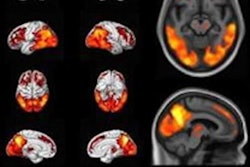
Given that clots contain an abundance of activated white blood cells and platelets, which are highly glycolytic, several studies noted the potential for FDG-PET/CT for detecting arterial and venous thrombosis in hospitalized COVID-19 patients, Gholamrezanezhad stated. In addition, one study noted decreased metabolic activity in the orbitofrontal cortex associated with COVID-19 induced anosmia.
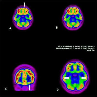
 FDG-PET/CT scan of a 27-year-old woman with persistent anosmia and positive PCR test for SARS-CoV-2. Representative axial (A, B, D) and coronal (C) images are shown. There was decreased uptake in the left orbitofrontal cortex (arrows). The uptake of temporal lobes were symmetric and normal (arrows, D). Image courtesy of Academic Radiology.
FDG-PET/CT and COVID-19 vaccination
More recent research has shown that reactive lymph node uptake on FDG-PET/CT may appear within a few days and persist for several weeks post COVID-19 vaccination.
"It is imperative that interpreting physicians recognize this manifestation in oncologic patients to avoid unnecessary biopsy and restaging," wrote Gholamrezanezhad.
In summary, Gholamrezanezhad and colleagues suggested that while FDG-PET/CT is usually not a first-line imaging modality, it can be a useful adjunct to traditional imaging techniques to help reduce COVID-19 disease burden and provide vital information at the molecular level.
"With the growing evidence of the detection of COVID-19 on FDG-PET/CT studies, radiologists and nuclear medicine physicians should be aware of the features of SARS-CoV-2 infection on FDG-PET/CTs, as this may lead to the detection of crucial non-oncologic pathologies," they concluded.