Researchers from the U.S. National Institutes of Health have developed a small, 3D-printable device for portable chest x-ray systems that improves estimates of head-of-bed (HOB) angle, according to a talk during a May 26 session of the Society for Imaging Informatics in Medicine (SIIM) annual meeting.
The patented device, called an X-clometer, could help radiologists interpret findings on x-rays such as pneumonia or other conditions involving pleural effusion. The X-clometer has been made available during the COVID-19 pandemic for use in clinical, academic, and research settings, according to Dr. Raisa Freidlin, an NIH staff scientist who presented the work at SIIM 2021.
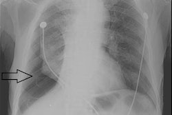
Portable chest x-rays are performed daily in most intensive care units at bedside when patients are too unstable to be transported to radiology departments for dedicated upright radiographs. For effective imaging, radiology technicians try to position patients at an upright angle, which lets fluid settle due to gravity and allows physicians to differentiate effusions from consolidations found in pneumonia, for instance. Patient positions are noted with lead arrows indicating they were either supine, semi-erect, erect, or upright at the time of the x-ray.
Moreover, radiologists cannot rule out free intraperitoneal air without knowing whether the image was acquired when the patient was in an erect position, nor can fluid levels such as hydropneumothorax be assessed consistently over time when patient positions vary.
"This compromised quality has been recognized for years. However, little has been done to provide a reliable indicator to optimize comparison of serial exams," according to Freidlin.
The researchers set out with the idea that if images can be obtained at similar angles each day, even if patients were not fully upright, a system could allow accurate comparisons and assessment of change. The first prototype was built in 2012 and back then was called UpRite, but it has been recently reduced in size and its accuracy has been improved.
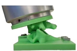
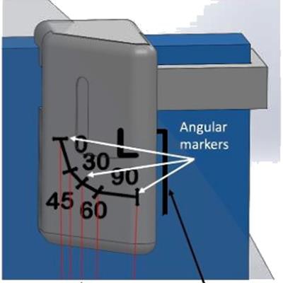
Operation of the X-clometer involves the x-ray cassette, the location of the X-clometer relative to the x-ray source, and divergent x-rays. The device is placed in the upper right corner of the field of view. Essentially, it is a left or right marker that also quantifies the head-of-bed angle from supine (0 degrees) to upright (90 degrees). The approximate angle is determined by a ball bearing that rolls freely within a curved passageway of the device to indicate the angle of the patient, cassette, and x-ray tube during the chest x-ray.
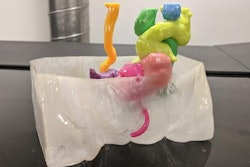
 The X-clometer. FOV = field of view. Image courtesy of Dr. Raisa Freidlin.
The system was created using both 3D model assembly programs Solidworks and Fusion 360. Using 3D computer-aided detection software, the researchers recently improved reading accuracy in the 60- to 90-degree range, which also reduced the size and optimized the positioning of the device. Since the device is smaller, less 3D printing material is required.
"We believe that knowing the degree of inclination across serial exams will help negate the need to bring patients to the department with numerous chest drains, IV lines, and other support devices," Freidlin concluded.
In addition, following further evaluation and actual use, X-clometer may decrease the need for obtaining CT scans, which would reduce unnecessary radiation exposure and additional expenses, she added.
The X-clometer 3D print files are publicly available on the NIH's 3D Print Exchange website.