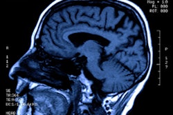
A new MRI technique called 3D amplified MRI (3D aMRI) can visualize brain motion in real-time, offering a noninvasive way to assess and track brain disorders, according to two studies published May 5 in Magnetic Resonance in Medicine and Brain Multiphysics.
The technique could be a powerful diagnostic tool for identifying hidden brain conditions before they become life-threatening, noted a team of researchers led by graduate student Itamar Terem of Stanford University in the Magnetic Resonance in Medicine paper. Terem's group included colleagues from Mātai Medical Research Institute in Gisborne-Tairawhiti, New Zealand; the Stevens Institute of Technology in Hoboken, NJ; and the University of Auckland.
"The new method magnifies microscopic rhythmic pulsations of the brain as the heart beats to allow the visualization of minute piston-like movements, that are less than the width of a human hair," Terem said in a statement released by the Stevens Institute. "The new 3D version provides a larger magnification factor, which gives us better visibility of brain motion, and better accuracy."
In the first paper, Terem and colleagues described the 3D aMRI method, comparing it with a 2D aMRI predecessor. In the second paper, a team led by Stevens Institute graduate student Javid Abderezaei outlined how the technique "visualizes, validates, and quantifies the amplitude and direction of the brain as it moves in three-dimensional space."
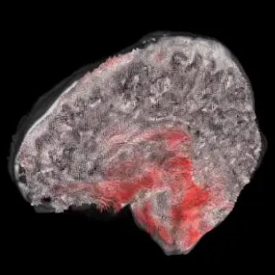
The 2D aMRI technique was developed by Terem; Samantha Holdsworth, PhD, director of research at Mātai; Mahdi Salmani Rahimi, PhD, also of Stanford; and other colleagues. The 3D technique builds on this previous work, using a video motion-processing method created by researchers from the Massachusetts Institute of Technology (MIT). It visualizes small movements of the brain at 1.2 mm3, the width of a human hair; these movements are then amplified up to 25 times so that clinicians can see them clearly. The study authors hope that the technique could offer more data on brain disorders such as Chiari I malformation, hydrocephalus, and aneurysms.
"We showed that 3D aMRI can be used for the quantification of intrinsic brain motion in 3D, which implies that 3D aMRI holds great potential to be used as a clinical tool by radiologists and clinicians to complement decision making for the patient's treatment," said the senior author of the Brain Multiphysics paper, Mehmet Kurt, PhD, of the Stevens Institute.
The investigators are using the technique to assess the effect of mild traumatic brain injury and are exploring, for example, whether the combination of 3D aMRI and brain modeling could lead to a noninvasive way of measuring brain pressure, which could help patients avoid surgery.
"What is exciting to see is that the dominant displacement patterns in the healthy brain qualitatively matched with the underlying physiology, which means that any changes in the physiological flow as a result of a brain disorder should be reflected in the displacements we measure," Abderezaei said in the statement.