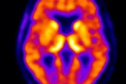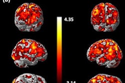
A new PET tracer may be effective for visualizing early pathological signs of Alzheimer's disease, according to a study by Swedish researchers that was published April 23 in Molecular Psychiatry.
Researchers at the Karolinska Institute in Stockholm studied the experimental radioligand BU99008 in normal tissue compared to brain tissue from six people who had died of Alzheimer's disease. They found the PET tracer could effectively detect a key receptor on cells called astrocytes, which are thought to play an early role in the development of Alzheimer's disease.
"Our study shows that BU99008 can detect important reactive astrocytes with good selectivity and specificity, making it a potentially important clinical astrocytic PET tracer," said lead author Amit Kumar, PhD, in a statement from the Karolinska Institute. "The results can improve our knowledge of the role played by reactive astrogliosis in Alzheimer's disease."
Reactive astrogliosis is the overproliferation of glial cells called astrocytes due to damage of nearby neurons from trauma, infection, or neurodegeneration, for instance. Several studies suggest that reactive astrogliosis may actually precede other known early pathological signs of Alzheimer's disease, including amyloid plaque and tau tangles. By identifying these cells with PET tracers, researchers hope to visualize their involvement in the development of the neurodegenerative disease.
BU99008 appeared in vivo in 2012 as a potential PET radioligand for imaging and has been led since through animal studies (pigs, rhesus monkeys, and Zucker rats) and a human dosing study by groups at Imperial College London and the National Institute of Radiological Sciences in Chiba, Japan.
In this study, the first in human samples, the Swiss researchers used brain tissue supplied by the Indiana University School of Medicine in Indianapolis from six individuals who had died with Alzheimer's disease and seven healthy controls who had died of other causes. They performed regional binding studies between reactive astrocytes and BU99008 on samples of the frontal, parietal, and temporal cortices; cerebellum; and hippocampus.
In normal tissue, the maximum binding (Bmax) value, which measures the number of binding sites per cell, was 58.8 femtomoles per milligram (fmol/mg). In the Alzheimer's disease samples, the Bmax for BU99008 was 316.2 fmol/mg. In other words, the experimental radiotracer showed a fivefold higher binding capability in Alzheimer's disease brain tissue than normal tissue, the authors stated.
"We also compared the regional distribution of 3H-BU99008 binding in [control] and [Alzheimer's disease] brain tissue homogenates and demonstrated significantly more extensive 3H-BU99008 binding in the frontal cortex (p = 0.0002) and hippocampus (p < 0.05) of the [Alzheimer's disease] brains as compared to [control] brains," Kumar and colleagues wrote.
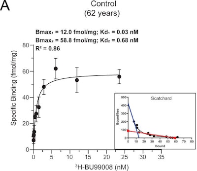 3H-BU99008 saturation binding studies were performed in temporal cortex brain tissue homogenates from one control and one patient with Alzheimer's disease (AD) using increasing concentrations of 3H-BU99008 (0-35 nM). A and B show the saturation binding curves for the control subject (62 years old) and the one with Alzheimer's disease (85 years old), along with the corresponding Scatchard plots. For the control, the second binding site was drawn manually after fitting the data using the Hill slope-specific binding function in GraphPad Prism 8.3 software (A; blue dotted line). Data are presented as means ± standard error of mean (SEM) of two experiments in triplicate. Kd = dissociation constant. Images courtesy of Molecular Psychiatry.
The authors noted some limitations to the study. The binding studies were performed on a small number of brains, and it would be interesting to further explore the BU99008 binding characteristics in larger sample sizes, they stated.
Nonetheless, this is the first time that BU99008 has been used to visualize reactive astrogliosis in Alzheimer's disease brain, the researchers noted.
"Further in vivo and in vitro studies in large numbers of [control] and sporadic and autosomal dominant [Alzheimer's disease] cases are needed to establish the reliability of BU99008 as a specific PET-tracer," they concluded.


