Deep learning-based image reconstruction software yields more accurate measurements of pulmonary nodules on low-dose chest CT exams than conventional reconstruction, according to a talk at the annual American Roentgen Ray Society (ARRS) meeting.
Researchers from South Korea compared two deep learning-based image reconstruction algorithms to a traditional hybrid iterative reconstruction technique in a phantom study encompassing four different radiation dose levels. Overall, they found that images produced by the two deep-learning algorithms led to significantly lower measurement error rates for lung nodules at every dose level, including an extremely low-dose setting comparable to a chest radiograph.
"Image noise was also significantly lower in [the deep learning-reconstructed images] compared to conventional [hybrid iterative reconstruction] at all radiation doses, including the extreme low-dose setting," said presenter Thomas Kwack of Korea University Ansan Hospital.
In 2011, the National Lung Screening Trial demonstrated that annual low-dose chest CT screening reduced relative mortality by 20% in individuals at high risk for lung cancer. Despite these advantages, repeated CT scans also pose the problem of radiation exposure, Kwack said.
Methods such as iterative reconstruction techniques have been developed to compensate for the increased noise that often accompanies lower dose on CT images. For example, the use of hybrid iterative reconstruction techniques has been shown to reduce image noise or artifacts and provides improved image quality over filtered back propagation in low-dose chest CT, according to Kwack. However, a recent study published in 2020 in European Radiology reported that deep learning-based image reconstruction could offer even better performance.
In their study, the South Korean researchers sought to investigate the accuracy of two commercial deep learning-based reconstruction software applications for enabling volumetry in subsolid and solid lung nodules. Specifically, they wanted to investigate the accuracy of these software applications in standard dose, low-dose, and extremely low-dose CT settings.
They first placed 10 synthetic nodules, including four types of solid nodules (3, 5, 8, and 10 mm in size) and three pure ground-glass nodules (5, 8, and 10 mm in size), into an anthropomorphic chest phantom. CT scans were then acquired at four different radiation dose settings: 120 kVp and 220 mA; 120 kVp and 90 mA; 120 kVp and 40 mA; and 80 kVp and 40 mA. The lowest-dose protocol produced an effective dose of 0.12 mSv, the equivalent of a chest x-ray, Kwack said.
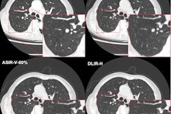
Images were reconstructed with three different reconstruction protocols: adaptive statistical iterative reconstruction (ASIR) from GE Healthcare and two deep learning-based reconstruction software applications: TrueFidelity (GE) and ClariCT.AI (ClariPi). Next, two radiologists independently measured nodule volumes in a semi-automated manner using version 4.4.12 of the Aquarius iNtuition edition software (TeraRecon).
The researchers then calculated the mean absolute percentage measurement error for the volume measurements from each type of reconstructed image. A smaller absolute percentage error indicates higher accuracy. They also calculated image noise to assess image quality.
| Mean volumetry absolute percentage error for all nodules on deep learning-reconstructed chest CT | |||
| ASIR | TrueFidelity | ClariCT.AI | |
| 120 kVp / 220mA | 7.27 ± 1.25 | 4.70 ± 0.50 | 3.09 ± 0.77* |
| 120 kVp / 90 mA | 9.87 ± 1.13 | 5.89 ± 1.38* | 5.95 ± 0.65* |
| 120 kVp / 40 mA | 12.25 ± 0.89 | 5.41 ± 1.00* | 7.72 ±1.90 |
| 80 kVp / 40 mA | 16.79 ± 1.75 | 6.99 ± 2.23* | 6.89 ± 2.24* |
"The [deep-learning] algorithms] showed significantly lower [absolute percentage error] than [ASIR] in every radiation dose setting," Kwack said.
Delving further into the data, the researchers found that both algorithms also produced significantly lower absolute percentage error than ASIR for 3-mm solid nodules across all dose levels. Absolute percentage errors were also significantly lower for nearly all ground glass nodules, especially at the lower dose settings, he said.
What's more, image noise was also significantly lower on both deep-learning algorithms than with the ASIR method at all radiation doses, Kwack said.