An artificial intelligence (AI) algorithm shows promise for helping clinicians triage abnormal brain MRI results, according to a study published April 21 in Radiology: Artificial Intelligence.
The findings could translate to better patient care, wrote a team led by Romane Gauriau, PhD, of Massachusetts General Hospital in Boston.
"Say you fell and hit your head, then went to the hospital and [staff] ordered a brain MRI," Gauriau said in a statement released by the RSNA. "This algorithm could detect if you have brain injury from the fall, but it may also detect an unexpected finding, such as a brain tumor. Having that ability could really help improve patient care."
As the use of brain MRI to evaluate brain injury has increased, radiology workflow has gotten more impacted, Gauriau and colleagues noted. Making use of AI to streamline the identification of abnormal findings in these exams could not only speed patient treatment but also reduce emergency room or hospital stays.
"There are an increasing number of MRIs that are performed, not only in the hospital but also for outpatients, so there is a real need to improve radiology workflow," Gauriau said. "One way of doing that is to automate some of the process and also help the radiologist prioritize the different exams."
The researchers collaborated with Diagnosticos da America SA (DASA) of Barueri, Brazil, to develop a deep-learning algorithm that uses T2-weighted fluid attenuated inversion-recovery (FLAIR) images to categorize brain MRI scans as either "likely normal" or "likely abnormal."
They trained the algorithm on three datasets that totaled more than 9,000 exams; each of the datasets were mined for data to create test sets.
- Model A (trained on dataset A, which was used to form test A),
- Model B (trained on dataset B, which was used to form test B), and
- Model A+B (trained on datasets A and B, which was used to form test C).
The researchers found sensitivity of up to 91% and specificity of up to 77% for abnormal brain MRI results, depending on the model and the test set compared with radiologist reports.
| Performance of AI algorithms compared with radiology report annotations | |||
| Measure | Model A | Model B | Model A+B |
| Sensitivity | |||
| Test A | 78% | 91% | 86% |
| Test B | 52% | 74% | 81% |
| Test C | 51% | 79% | 66% |
| Specificity | |||
| Test A | 69% | 32% | 57% |
| Test B | 77% | 72% | 72% |
| Test C | 74% | 52% | 69% |
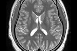
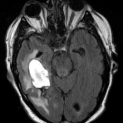
 Examples of axial FLAIR sequences from studies within dataset A. From left to right: a patient with a "likely normal" brain, a patient presenting an intraparenchymal hemorrhage within the right temporal lobe, a patient presenting an acute infarct of the inferior division of the right middle cerebral artery, and a patient with known neurocysticercosis presenting a rounded cystic lesion in the left middle frontal gyrus. Image and caption courtesy of RSNA.
"[Our study shows] promising results ... when comparing model performance to annotations from the radiologic reports, indicating that such an approach could be used to build a triage system for MRI brain examinations," the group noted.
As for further research, Gauriau and colleagues plan to further develop the algorithm beyond the "likely normal" and "likely abnormal" categories.
"This way we could not only have binary results but maybe something to better characterize the types of findings, for instance, if the abnormality is more likely to be related to tumor or to inflammation," she said.