A new 3D imaging technique that improves brain tumor visibility on MRI has been described by researchers from Northwestern University in Chicago in a study published October 28 in Science Advances.
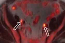
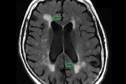
Called T1 relaxation-enhanced steady-state (T1RESS), the new technique applies radio waves and magnetic fields that generate MRI signals differently from current methods, producing improved tumor visibility. It is particularly useful for identifying very small malignant lesions, lead author Dr. Robert Edelman said in a statement released by the university.
"For patients undergoing treatment by surgery or radiotherapy, it is hoped the improved visibility of the tumor margins on contrast-enhanced scans will ensure the entire tumor is treated and result in better outcomes," he said.
The investigators' study included 54 patients with brain tumors and showed that T1RESS improved the contrast between tumors and normal brain tissue two-fold compared with existing MRI techniques. Edelman likens T1RESS's effect to seeing the stars at night rather than trying to see them during the day.
"There just isn't enough contrast between the stars and the sunlit sky to make them visible," he said. "In the case of brain tumors, T1RESS doubles the contrast between tumors and normal brain, so the tumors are more easily detected. It's like looking at the stars on a dark night instead of on a sunny day."
If the benefits of T1RESS are confirmed in further studies, it would be relatively easy to make it available because it consists of a specialized software package, according to the group. The researchers plan to investigate the use of the technique with breast and prostate cancer as well, the university said.
Edelman filed for a patent for T1RESS earlier this year with the U.S. Patent and Trademark Office, and a notice of allowance was issued by the office in September.