A new artificial intelligence (AI) calculator for analyzing brain tissue damage derived from MRI scans can detect with 70% accuracy early signs of cognitive decline, according to a study published October 27 in Academic Radiology.
The algorithm suggests a better way to assess small bright spots on MRI called white-matter hyperintensities, according to a team led by senior author Dr. Yulin Ge of NYU Langone Health in New York City. The spots are fluid-filled holes in the brain that develop from the breakdown of the blood vessels that support nerve cells and are linked to memory loss and emotional problems as people age.
"Our new calculator for properly sizing white-matter hyperintensities, which we call bilateral distancing, offers radiologists and other clinicians an additional standardized test for assessing these lesions in the brain, well before severe dementia or stroke damage," Ge said.
White-matter hyperintensities are easily imaged on MRI, but their origin and pathophysiology aren't always clear, the group noted. Typically, radiologists assess these lesions based on visual interpretation and a three-point scale. The new calculator offers an objective way to evaluate the volume and location of the lesions, according to the researchers.
For the study, lead author Jinyun Chen, PhD, Ge, and colleagues included 60 MR scans from the Alzheimer's Disease Neuroimaging Initiative (ADNI) of both elderly men and women mostly over the age of 70 with normal brain function, mild cognitive decline, or severe dementia (20 exams for each group). The group developed an algorithm that calculated lesion volumes based on distance from both sides of the brain, with normal ranges being between 0 and 60 milliliters. Volumes of more than 100 milliliters indicated severe disease.
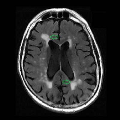
 MRI image of human brain shows multiple bright spots (white-matter hyperintensities) in center (as shown by green arrows). Images and caption courtesy of Academic Radiology.
The group checked the measurements generated by the algorithm against the MRI findings and found that seven out of 10 matched the patient's diagnosis.
"Amounts of white-matter lesions above the normal range should serve as an early warning sign for patients and physicians," Chen said in the NYU statement.
While white-matter brain lesion measures alone aren't enough to certify a finding of early dementia, they should be taken into consideration along with factors such as a history of brain injury, memory loss, hypertension, or clear signs of cognitive decline.
In any case, the tool should help clinicians better monitor their patients' brain health, according to the team.
"With a standardized tracking and measuring tool, it is now possible to monitor the growth of white-matter lesions relative to that of other tau and beta-amyloid proteins also believed to be potential causes of dementia and Alzheimer's disease," the university said. "The buildup of either substance could also prove or disprove one or more of the theories about what biological processes actually lead to various forms of dementia."
Chen's group plans to further test the calculator with another 1,495 brain MRI scans, according to NYU Langone Health.