Researchers from Massachusetts General Hospital (MGH) have validated a new radiolabeled molecule that can be used with imaging tests to accurately detect and characterize brain injury. The U.S. Food and Drug Administration (FDA) recently cleared the study team, led by Nicolas Guehl, PhD, an investigator in the department of radiology at MGH, to begin a first-in-human study.
The tracer, called F-18 3F4AP, is designed to bind to potassium channels and is radiolabeled so it can be visualized with PET. Potassium channels become exposed when the brain's neurons are demyelinated, which occurs with a variety of neurodegenerative conditions such as multiple sclerosis and Alzheimer's disease.
During an animal test involving monkeys, the tracer had high penetration into the brain, fast washout, and was reproducible. F-18 3F4AP also showed unusually high resistance to metabolic degradation and minimal binding to blood proteins, according to Guehl, who is also an instructor in radiology at Harvard Medical School.
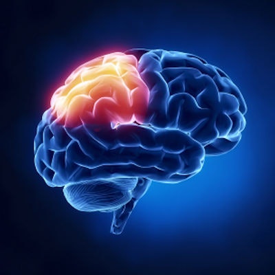
In addition, F-18 3F4AP generated a high signal in the right frontal cortex of a monkey that the researchers later learned had sustained a minor brain injury several years before being transferred to them.
Human testing will begin imminently, according to the researchers.