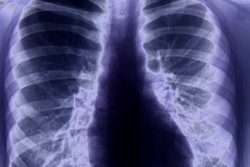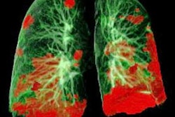
Researchers have created computerized models of lung abnormalities found on CT scans of patients with COVID-19, according to an August 28 study in the American Journal of Roentgenology. The virtual imaging models can be used like computerized phantoms for research into the best use of imaging to detect and monitor the disease.
After manually segmenting morphologic features of COVID-19 abnormalities from 20 CT scans, a team of researchers led by Ehsan Abadi, PhD, of Duke University created digital images that reflected the size and shape of the imaging manifestations. These models were then incorporated into an adult male 4D extended cardiac-torso (XCAT) phantom developed by Duke; the texture and material of the lung parenchyma in abnormal areas were modified in the phantom to match the properties shown on the clinical images.
"The developed toolsets in this study provide the foundation for use of virtual imaging trials in effective assessment and optimization of CT and radiography acquisitions and analysis tools to help manage the COVID-19 pandemic," the researchers wrote.
Abadi and colleagues created three COVID-19 computational phantoms and then virtually imaged the models using a Definition Flash CT scanner (Siemens Healthineers) and a radiography simulator (DukeSim).
The simulated abnormalities -- ground-glass opacities and consolidation -- were visible and realistic in terms of shape and texture on both simulated CT and radiographic images, according to the authors. The team also found that contrast-to-noise ratios in the abnormal regions of the image were 1.6 for 5 mAs images, 3 for 25 mAs images, and 3.6 for 50 mAs images.
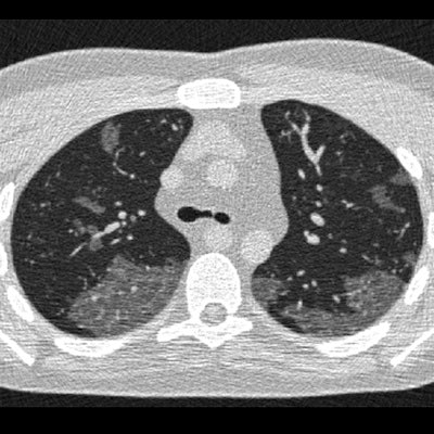
 Simulated images of COVID-19 on 4D extended cardiac-torso phantom developed at Duke University, including simulated CT at 50 mAs (A), 25 mAs (B), and 5 mAs (C), as well as a simulated chest radiograph (D). Images and caption courtesy of the American Journal of Roentgenology.
With virtual imaging trials, researchers could potentially investigate and optimize the role of radiography and CT in diagnosis and follow-up of COVID-19. These techniques enable the quality and performance of these imaging modalities to be comprehensively assessed and compared, according to Abadi et al. What's more, radiation dose and image quality could be quantitatively optimized.
"Because virtual imaging trials have predefined models of the disease, they can provide a valuable tool to train and evaluate either principle-informed or machine-learning algorithms designed for disease classification and quantification," the authors added.
The researchers are now building on their framework by modeling a larger population of COVID-19 XCAT phantoms using more CT exams from patients. These phantoms will be informed by CT images acquired at different disease stages, as well as clinical information and longitudinal studies, according to the authors.
"It is crucial to have these models at different stages of the disease because they are envisioned to be used for various imaging studies from screening to treatment evaluations and follow-up care," they wrote.
In addition, Abadi et al said they are now performing systematic simulation of imaging protocols and task-based characterizations of the resulting images.
"With these tools, we will perform virtual imaging trials to compare radiography and CT methods and protocols with the goal of defining COVID-19 protocols for optimized screening and follow-up of patients, with the confidence of knowing the expected performance," they wrote.
Potential applications include comparing COVID-19 screening performance for chest radiography and CT, as well as determining the effects of radiation dose on detection accuracy and further optimizing this parameter in imaging COVID-19 patients, according to the authors.
"Our virtual toolsets can also be used to optimize the imaging and analysis tools in terms of sensitivity and specificity for the detection and classification of the disease," they wrote.