When it comes to abbreviated breast MRI, what's the best image acquisition protocol? Researchers from Minnesota tested diffusion-weighted imaging (DWI) techniques to come up with the most optimal method and published their results August 25 in Radiology.
Researchers from the University of Minnesota found that a five-minute DWI-MRI protocol that leveraged an axillary-reformatted simultaneous multislice (AR-SMS) technique showed significantly better performance than read-segmented and spin-echo DWI-MRI. The use of alternative, high-spatial-resolution protocols, including AR-SMS, could help make abbreviated MRI more feasible for everyday breast imaging.
"For breast diffusion-weighted imaging, axially reformatted-simultaneous multislice imaging provided the highest spatial resolution, covered the largest volume, and helped to detect the smallest feature compared with readout-segmented echo-planar imaging and standard spin-echo echo-planar imaging," wrote the authors, led by Jessica McKay, PhD, and her colleagues at the University of Minnesota Center for Magnetic Resonance Research.
MRI has shown promise as a breast imaging technique for decades, but long image acquisition times have hindered more rapid adoption, particularly for screening. Recently, faster MRI acquisitions -- called abbreviated breast MRI -- offer the promise of making MRI more economical as a screening tool.
Spin-echo echo-planar imaging (EPI) is considered the standard DWI-MRI protocol, and it has been investigated for screening and disease characterization, as well as for monitoring response to treatment.
But spin-echo DWI has drawbacks that are particularly problematic for breast imaging, including low spatial resolution, large geometric distortions, and artifacts stemming from patient motion. New protocols with higher spatial resolution, including AR-SMS and read-segmented echo-planar imaging, have been proposed as potential alternatives for breast imaging.
In their prospective study, the authors compared AR-SMS, read-segmented echo-planar, and spin-echo echo-planar protocols in a sample of 30 lesion images acquired from women undergoing DWI-MRI for breast screening or diagnostics at the University of Minnesota.
The protocols were optimized for a five-minute acquisition time using a 3-tesla MRI scanner with a 16-channel breast coil. The authors used commercially available software to reconstruct the spin-echo and read-segmented DWI-MRI images, but they used a custom Matlab script for the AR-SMS images.
Three blinded radiologists interpreted the images, measuring the longest dimension on contrast-enhanced subtraction images and images with b values of 800 sec/mm2. They also used a five-point scale to rate overall image quality and their confidence in their measurements.
All three radiologists ranked AR-SMS highest for overall image quality, and the protocol received significantly better image quality marks than the other two methods. On the five-point scale, AR-SMS images scored 1.31 points higher than spin-echo echo-planar images and 0.74 points higher than read-segmented echo-planar images.
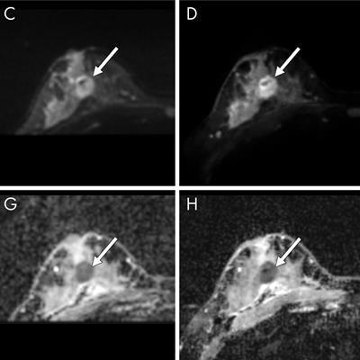
 Images of a large lesion from a clinical full-protocol MRI (clinical MRI), standard single-shot spin-echo echo-planar imaging (Standard A), readout-segmented echo-planar imaging (RS-EPI), and axially reformatted-simultaneous multislice imaging (AR-SMS). The images in the clinical MRI column include (A) contrast-enhanced image and (E) T2-weighted image. The images in the alternative MRI protocol columns include (B-D) images with b value of 800 sec/mm2 and (F-H) apparent diffusion coefficient maps focused on a large contrast-enhanced lesion. Image courtesy of the RSNA.
The radiologists also expressed more confidence in lesion size measurements taken from the high-spatial DWI-MRI protocols. AR-SMS and read-segmented EPI both scored significantly better than spin-echo EPI on the five-point scale.
While AR-SMS often came up on top in the analysis, read-segmented EPI still held its own. Read-segmented EPI had significantly better image quality scores than standard spin-echo EPI, and the difference between read-segmented echo-planar images and AR-SMS was not significantly different for confidence in lesion size measurements.
Notably, AR-SMS has the advantage of providing full-coverage but also requires extensive distortion correction, while read-segmented EPI is available on commercial systems but has limited use for visualizing anterior-posterior regions.
The authors hypothesized that future protocols may be able to combine the strengths of the two methods to create a new DWI-MRI protocol that is better than the sum of its parts.
"Combining the encoding speed of AR-SMS with the reduced distortion of [read segmented] echo-planar imaging may be a promising strategy for future work," the authors wrote.