A new PET radiotracer may one day help clinicians evaluate stroke recovery, according to research presented at the Society of Nuclear Medicine and Molecular Imaging (SNMMI) 2020 virtual annual meeting. Researchers used the radiotracer, F-18 SynVesT-2, to assess synaptic density in rat brains.
Current stroke imaging with F-18 FDG PET relies on measurements of oxygen or glucose metabolism. But transient hyperglycemia -- a common occurrence in stroke patients -- can interfere with FDG uptake and, consequently, the findings of an F-18 FDG-PET scan.
To overcome this limitation, researchers from Yale University developed a new imaging method that directly visualizes synapses. Synapses are not only important for brain functions, but a synaptic deficit is also a hallmark sign of stroke, noted lead author Xueying Lyu, a postgraduate research associate at the Yale School of Medicine.
"This is the first direct demonstration of synaptic changes following stroke and has shown that the synaptic protein SV2A is a potential biomarker for tissue viability," Lyu stated in a press release.
To test the radiotracer, the researchers performed weekly F-18 SynVesT-2 PET scans on six rats with simulated strokes. They acquired four scans in total, starting the first day after simulated stroke incidence, and used the scans to conduct image analysis and generate standardized uptake values (SUVs) for different parts of the brain.
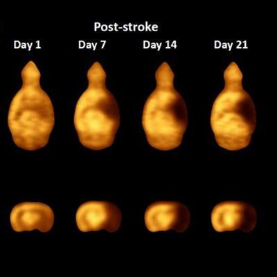
 Average SUV ratio PET images of stroke rat model over time as compared with a sham rat using mean cerebellum as the reference. Image courtesy of T. Toyonaga and X. Lyu, Yale University, New Haven CT.
The new imaging method enabled the researchers to successfully detect synapse loss in the rats. The loss of synapses was most evident in the hippocampus, thalamus, and neocortex, but not the cerebellum.
The researchers were also able to track disease progression through lesion quantification. The most significant synaptic loss occurred in the first week after stroke, the researchers noted.
The new imaging method is preliminary, but it could one day help with stroke detection and recovery monitoring in acute stroke patients. Lyu, in particular, is optimistic about the technology reaching clinical settings.
"Information on tissue viability is valuable to detect stroke early and to evaluate therapeutic efficacy and recovery," he stated. "We hope to help stroke patients by bringing this SV2A PET imaging method to clinical settings in the near future."