MRI scans of 11 astronauts performed the day after their return from extended time in space have revealed significant changes in brain volume and cerebrospinal fluid (CSF) flow. The astronauts also saw their pituitary glands shrink, according to a study published April 14 in Radiology.
In fact, the greater amount of white-matter volume and elevated velocity of CSF through the cerebral aqueduct -- the narrow channel that connects the ventricles in the brain -- remained steady one year after the astronauts returned to Earth.
"What we identified that no one has really identified before is that there is a significant increase of volume in the brain's white matter from preflight to postflight," said lead author Dr. Larry Kramer from the University of Texas Health Science Center in Houston, in a statement. "White-matter expansion in fact is responsible for the largest increase in combined brain and cerebrospinal fluid volumes postflight."
These findings are not the first time abnormalities have been found in astronauts who spent long periods of time away from the Earth. As far back as March 2012, Kramer and colleagues reported optical abnormalities in the eyes and brains of 27 astronauts who underwent MRI scans after exposure to microgravity or zero gravity for an average of 108 days while on space shuttle missions and/or serving on the International Space Station.
By November 2017, Roberts and colleagues reported their MR images showed a narrowing of CSF spaces and the central sulcus. More recently, an October 2018 study by Eulenburg et al found the loss of gray-matter volume on MR brain images soon after touchdown and prolonged changes in circulation of CSF seven months later.
In the current study, Kramer and colleagues prospectively enrolled 11 astronauts (mean age, 45 ± 5 years) who voluntarily underwent 3-tesla MRI scans (Verio, Siemens Healthineers) with a 32-channel head coil before blastoff and one, 30, 90, 180, and 360 days after their return to Earth. During those scans, the researchers measured white- and gray-matter volume, CSF volumes, and CSF hydrodynamics, which gauge the levels of peak CSF blood flow velocity in the brain. The astronauts' mean exposure time to microgravity was 171 (± 71) days.
When the researchers compared MR images before time in space and one day after the astronauts' return, they discovered a number of statistically significant changes, including the following:
- Increased mean total volume in the brain of 28 mL (p < 0. 001), most of which was due to white matter of 26 mL (p < 0.001)
- Increased mean enlargement of lateral ventricles of 2.2 mL (p < 0001)
- Mean CSF peak-to-peak velocity magnitude increases 2.2 cm/sec (p = 0.01)
- No significant changes in the brain's gray matter.
"When you're in microgravity, fluid such as your venous blood no longer pools toward your lower extremities but redistributes headward," Kramer said in a statement. "That movement of fluid toward your head may be one of the mechanisms causing changes we are observing in the eye and intracranial compartment."
When brain volume and CSF were combined, the statistically significant increases held steady for one year. In addition, six astronauts (55%) showed a significant decrease in the mean pituitary gland of 5.3 mm, compared with 5.9 mm in the preflight MRI (p < 0.01).
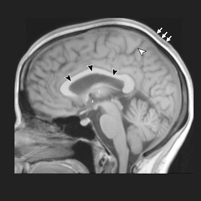
 Reconstructed 5-mm orthogonal midline MR brain images using the sagittal 3D T1-weighted dataset show preflight baseline image (a) and matching postflight image (b) one day after return in the same astronaut. Black arrowheads show upward expansion of the anterior, middle, and posterior superior margins of the lateral ventricle with associated narrowing of the marginal sulcus of the cingulate sulcus (white arrowhead). There also is subtle expansion of the third ventricle (indicated by a 3, middle of left image), which has displaced the thalamus (T, left image) from midline, making it less visible. There is thickening of the intermediate signal scalp soft tissues (arrows). Images courtesy of Radiology.
Fortunately for the space travelers, the aforementioned changes in brain volume, CSF, and the pituitary gland are not considered problems for healthy adults. The researchers liken the conditions to people with extended bed rest.
Kramer and colleagues are exploring ways to counteract the effects of microgravity and believe these findings can be used to help people who prefer their feet firmly planted on the ground.
"If we can better understand the mechanisms that cause ventricles to enlarge in astronauts and develop suitable countermeasures, then maybe some of these discoveries could benefit patients with normal pressure hydrocephalus and other related conditions," Kramer said.