A combination of chest x-ray and lung ultrasound offers a workable way to manage complicated pneumonia in children -- although MRI is a more effective modality when it comes to identifying particular features, according to a study published online March 19 in PLOS One.
The results suggest that, if MRI is not readily available, x-ray offers an alternative to the higher radiation a modality like CT would impart on follow-up, wrote a team led by Dr. Philip Konietzke of University Hospital of Heidelberg in Germany.
"MRI demonstrates to be more sensitive than chest x-ray in detecting abscess and empyema in complicated pneumonia, but it showed no advantages in detecting consolidation and pleural effusion," the group wrote. "MRI can contribute to a better clinical management of complicated pneumonia in hospitalized children and can potentially replace chest CT as a radiation-free modality ... [but] the availability of MRI scanning time is still limited, and the need for sedation as well as the administration of contrast media with its possible side effects has to be considered in noncooperative young children."
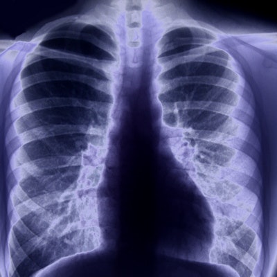
Chest x-ray has long been the first imaging modality for workup of children with pneumonia, and it is sometimes paired with lung ultrasound. If patients develop complications like abscesses or empyema, they often undergo more chest x-rays or CT exams. But this follow-up increases their exposure to radiation, which is why researchers have been seeking a radiation-free alternative, the authors noted.
MRI offers this alternative but may not be readily available. So Konietzke and colleagues conducted a study to investigate whether chest x-ray with lung ultrasound could perform comparably to MRI for evaluating the presence and severity of abscess, consolidation, bronchial wall thickening, mucus plugging, and pleural effusion in pneumonia. The study included 33 children (average age, 6 years) who had 33 paired MRI and chest x-ray scans. Both groups also received ultrasound.
Although MRI found more lung abscess formations compared with chest x-ray -- both alone and with lung ultrasound -- the combination of chest x-ray and ultrasound proved comparable to MRI when it came to identifying pleural effusion and empyema.
| Chest x-ray performance compared with MRI for identifying pneumonia in children | ||||||
| Findings | Chest x-ray | MRI | p-value | Chest x-ray plus lung ultrasound | MRI | p-value |
| Pulmonary abscess/necrosis formations | 27.3% | 72.7% | 0.001 | 55.6% | 77.8% | 0.109 |
| Consolidation | 97% | 100% | 1 | 96.3% | 100% | 1 |
| Bronchial wall thickening | 27.3% | 97% | 0.001 | 0% | 96.3% | 0.001 |
| Mucus plugging | 0% | 66.7% | 0.001 | 0% | 33% | 0.004 |
| Pleural effusion/empyema | 81.8% | 93.9% | 0.125 | 88.9% | 92.6% | 0.895 |
The study findings led the researchers to conclude that chest x-ray with lung ultrasound offers a workable way to diagnose and manage pneumonia in children, even though MRI performs better for some matrices.
"Chest x-ray and lung ultrasound seem to be sufficient in most cases," the team concluded. "In cases where lung ultrasound cannot be realized or the combination of chest x-ray and lung ultrasound might not be sufficient, MRI, as a radiation-free modality, should be preferred to CT."