What effect does vaping have on the lungs of young, healthy individuals? A study published online March 3 in Radiology details the most prevalent findings seen on the chest x-rays and CT scans of teenagers diagnosed with electronic cigarette (e-cigarette), or vaping, product use-associated lung injury (EVALI).
Since 2014, e-cigarettes have been the most widely used tobacco product among youths in the U.S., with increasing usage rates over the past several years. But vaping has recently been under fire for potentially provoking the sudden rise of an otherwise unexplainable severe respiratory illness. As of January 14, the number of EVALI cases reported to the U.S. Centers for Disease Control and Prevention (CDC) was 2,668, including 60 confirmed deaths.
Though the incidence rate of EVALI has gradually decreased since its peak in September 2019, occasional reports of vaping-related lung injury in teenagers, a particularly vulnerable population, continue to pose a concern.
As part of a larger investigation into EVALI, Dr. Maddy Artunduaga and colleagues from the University of Texas Southwestern Medical Center evaluated the imaging data of 14 teenagers with EVALI who underwent chest x-ray and CT exams. The average age of the patients was 16 years, and half were male. All of the patients had a history of vaping or dabbing, which involves inhaling small but highly concentrated doses of oil with THC, within 90 days before symptom onset.
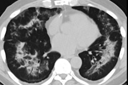
Chest x-rays showed ground-glass opacity in 100% and consolidation in 57% of the patients. The extent of abnormality on x-ray covered 50% to 75% of the lungs for about a third of the cases and more than 75% of the lungs in another third.
On the CT scans, ground-glass opacity was evident in all of the patients, consolidation in 64%, and interlobular septal thickening in 14%, with subpleural sparing in 79% of the cases. The extent of abnormality visible on CT exceeded 75% of the lungs in the vast majority of cases. About 36% of the patients' CT scans also showed the reversed halo sign.
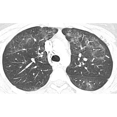
 Contrast-enhanced chest CT image of a 16-year-old girl with EVALI showing central ground-glass opacity surrounded by a dense ring of consolidation (arrows) as well as subpleural spacing (arrowheads). Image courtesy of the RSNA.
For both imaging modalities, the abnormalities were bilateral for all cases and were symmetric in all but one. Half of the x-rays and CT scans showed lower lobe predominance. Interobserver agreement was nearly perfect between the two pediatric radiologists who examined the chest x-rays and CT scans.
The researchers found that chest x-ray alone provided an insufficient characterization of EVALI patterns in pediatric patients, underscoring the need to include CT for adequate assessment.
"CT provides a better evaluation of the extent and degree of involvement, as well as a better assessment of the characteristics of abnormal pulmonary findings," Artunduaga said in a statement. "This can lead to the early diagnosis of EVALI, which would ultimately allow the timely management of this entity in the pediatric population."
Artunduaga and colleagues also noted that many young patients initially present with a wide range of symptoms -- including respiratory, gastrointestinal, and constitutional symptoms -- that may misdirect clinicians away from suspecting EVALI, which further emphasizes the importance of imaging in this population.
"Radiologists will continue to play an important role in the initial detection and follow-up of pediatric patients with EVALI," she said. "In some cases ... the radiologist may be the first to suggest EVALI as a diagnostic consideration."