U.S. and Chinese researchers have combined an advanced image reconstruction method with a total-body PET scanner to track real-time blood flow and heart and respiratory function, according to a study published online January 20 in the Proceedings of the National Academy of Sciences.
The video created by researchers from University of California, Davis (UCD) and Fudan University in Shanghai could become a way to simultaneously observe the function of multiple organs, such as the brain and the heart.
"The results show that we can visualize radiotracer transport in the body on timescales of 100 [msec] and obtain motion-frozen images with superior image quality compared to conventional methods," wrote the authors, led by Xuezhu Zhang, PhD, from the UCD department of biomedical engineering.
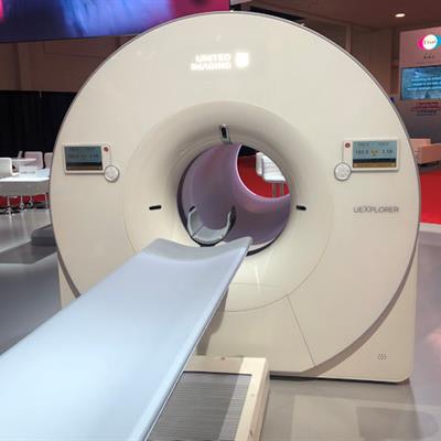
The researchers used the Explorer total-body PET/CT system, a 3D 194-cm-long hybrid scanner that was developed by researchers at UCD under the direction of Simon Cherry, PhD, professor of biomedical engineering at UCD, and Ramsey Badawi, PhD, professor of radiology at UCD School of Medicine.
A commercial version of the scanner, dubbed the uExplorer, currently is manufactured and marketed by United Imaging Healthcare of Shanghai. The device has received clearance from the U.S. Food and Drug Administration and is now in clinical use at UCD.
For this study, the researchers sought a way to perform ultrahigh (100 msec) temporal resolution dynamic PET imaging by combining advanced dynamic image reconstruction with the uExplorer. More specifically, the goal was to acquire the real-time activity of radiotracer distribution and uptake, as well as cardiac motion, by reducing noise and reconstructing images from the raw data of volunteers.
The resulting video shows the motion of the heart with exceptional clarity, with changes in the cardiac contraction visible with clear delineation of the end-systolic and end-diastolic phases.
"The breakthrough in this work is to capture the ultrafast whole-body dynamic tracer imaging with Explorer at the same time," said Jinyi Qi, PhD, professor of biomedical engineering at UCD, in a statement. "We can see global changes with improved image quality at a timescale of 100 [msec], which was never seen before using any medical imaging modalities."
Among the clinical benefits of this approach is the ability to measure cerebral blood flow and cerebral metabolic rate of oxygen, the authors noted.
"Furthermore, without the artifacts induced by cardiac and respiratory motion, ultrafast PET may allow analysis of metabolic processes within atherosclerotic plaques and evaluate their distribution and characteristics throughout the cardiovascular system," Zhang and colleagues wrote. "Finally, high temporal resolution PET together with the TB coverage allows dynamic tracer studies of brain-heart and brain-gut interaction."