A new optical technique can determine the differences between potentially life-threatening coronary plaques and plaques that pose less immediate danger to patients with heart disease, according to a first-in-human study.
A team of researchers from Massachusetts General Hospital (MGH) and the U.S. National Institute of Biomedical Imaging and Bioengineering (NIBIB) led by Brett Bouma, PhD, believe their new method could change the game for cardiologists by helping them better identify patients at higher risks of future heart attacks and helping them improve medical therapy, according to a release issued by NIBIB.
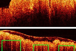
The researchers used intravascular polarimetry to investigate the polarization properties of coronary atherosclerotic plaques in 30 patients with coronary artery disease by searching for indications of plaque instability. Intravascular polarimetry is an optical technique used to assess the polarization properties of cross-sections of vascular tissue, according to their study (JACC: Cardiovascular Imaging, August 8, 2019).
The electric field of polarized light creates a wave signal along a single plane, and when it is directed at tissue, the electric field is influenced by the tissue's microscopic structure and organization. Tissues rich in collagen and smooth muscle cells split the beam of light into two rays that diverge into slightly different directions. The optical effect allowed MGH and NIBIB researchers to distinguish coronary plaque composition and stability, the release stated.
To gather the data, participants were required to undergo cardiac catheterizations, including intravascular imaging with optical coherence tomography. The catheterizations provided multiple plaque images for each procedure, including 342 cross-sectional plaque images and 244 images from the fibrous caps of the atherosclerotic lesions responsible for high-risk or stable symptoms.
The high-resolution images enabled the researchers to classify coronary cross-sections into one of seven categories: normal, fibrous, fatty, calcified, thick cap, thin cap, or ruptured cap. Then, a specialized instrument was used to determine the polarization properties of their coronary arterial walls, according to the authors.