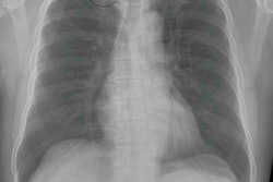
Digital variance angiography (DVA) uses an image processing algorithm to visualize contrast motion and calculate the variance of each pixel intensity in a series of x-ray images.
Dr. Viktor Orias from the Heart and Vascular Center of Semmelweis University Budapest will present the details of two observational studies that included 66 patients undergoing lower limb x-ray angiography due to symptomatic peripheral arterial disease.
Orias and colleagues compared the signal-to-noise (S/N) ratios of digital subtraction angiography (DSA) and DVA in a subgroup of 24 patients who received carbon dioxide (CO2) as a contrast agent and another group of patients who received iodinated contrast medium (ICM). A total of 6,814 regions of interest were selected on image pairs to compare S/N ratios from DVA and DSA.
In the iodinated contrast group, DVA provided an average 3.3 times higher S/N ratio than DSA on angiograms. In 69% of the iodinated contrast comparisons, the researchers found DVA provided higher-quality images than DSA. In addition, the S/N ratio was an average 3.0 times higher in DVA images with CO2.
"DVA provides significantly higher image quality than DSA while performing catheter angiography with ICM or CO2," Orias and colleagues wrote in their abstract. "The higher image quality makes CO2 angiography more feasible for patients with renal insufficiency and iodine allergy."
The significant S/N ratio difference also suggests it is possible to reduce radiation and ICM exposure, but additional study is needed to determine the optimal protocols for low-dose DVA acquisition, the researchers noted.