The findings with ultrahigh-field MRI and susceptibility-weighted imaging (SWI) could prove worthwhile, given the potential association between cerebral microbleeds and chronic traumatic encephalopathy (CTE).
Dr. Cornelius Deuschl, a radiologist at Essen University Hospital, and colleagues enrolled 12 professional football players for this prospective study. All subjects underwent 3-tesla (Magnetom Skyra, Siemens Healthineers) and 7-tesla (Magnetom 7T, Siemens) MRI scans with SWI and 32-channel radiofrequency head coils.
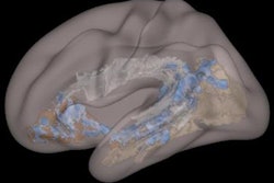
With 3-tesla SWI-MRI, two neuroradiologists spotted three cerebral microbleeds in three different players. At 7-tesla SWI, the readers confirmed one of the three cerebral microbleeds and two smaller adjacent cerebral microbleeds. As for the two suspected cerebral microbleeds on 3-tesla MRI, 7-tesla SWI correctly identified them as atypical small intracerebral veins.
Given 7-tesla SWI's accuracy in determining cerebral microbleeds, Deuschl and colleagues believe it can contribute to future knowledge on how repeated head trauma will affect football players and patients with similar injuries.