The group was inspired by the Pediatric Bone Age Challenge at RSNA 2017, which demonstrated machine learning's potential to accurately estimate quantifiable data, according to Presenter Dr. Peter Kamel of Johns Hopkins Hospital in Baltimore.
"[Our] project aimed to extend this type of quantitative machine learning by cross-training radiology images on [dual-energy x-ray absorptiometry] reports and provide accurate estimates of bone mineral density on radiography, which is currently very subjective from a radiologist standpoint," he told AuntMinnie.com. "Osteoporosis is a highly prevalent and highly impactful disease, and using machine learning in this regard has the opportunity for great clinical benefit."
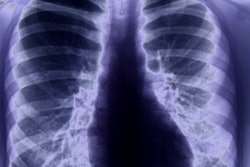
The researchers trained and tested a variety of deep convolutional neural networks on a dataset of 875 postmenopausal women who had received DEXA scans, as well as a posteroanterior and lateral chest radiographs within three months of the DEXA study. Their algorithm could distinguish osteoporotic and normal radiographs with an area under the curve of 0.87.
"We provide proof of concept that deep learning can be fit to accurately quantify information in medical imaging that is very subjective on human interpretation, and we do this by cross-training with labels from another modality," Kamel said. "Specifically, this application may provide the ability for opportunistic screening for osteoporosis on radiographs at no added risk to patients and may potentially be used for cases where DEXA scanning cannot be performed or is unavailable."
Get all the details by sitting in on this late-morning talk on Monday.