Using 7-tesla MRI and a custom-built head coil, researchers have produced an image of the human brain with 100-μm resolution and the kind of explicit detail that someday might be used to investigate neurological disorders, according to a study published October 30 in Scientific Data.
"This unique dataset has a broad range of investigational, educational, and clinical applications that will advance understanding of human brain anatomy in health and disease," said lead author Dr. Brian Edlow, the associate director of Massachusetts General Hospital's Center for Neurotechnology and Neurorecovery.
A donated brain specimen was scanned on a clinical whole-body 7-tesla system (Siemens Healthineers) with a specially designed radiofrequency device that features 32 coils positioned closely enough together to fit snugly around a human brain without interfering with each other's operation.
The goals of the researchers include producing an in-depth view of brain structure and the connections between its regions. By doing so, they hope to add to current knowledge on the biology of neurological disorders, such as traumatic brain injury and Alzheimer's disease, through a new type of "network-based autopsy" that integrates MRI with histopathology. If all goes well, these future ultrahigh-resolution imaging techniques potentially could be used with living subjects.
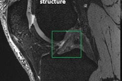
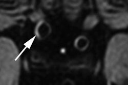
In one application, the researchers integrated data from the scan into a software platform called Lead DBS, which might allow neurologists and neurosurgeons to improve the therapeutic outcome of deep brain stimulation electrode placement in disorders that include Parkinson's disease and obsessive-compulsive disorder.
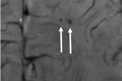
In addition, researchers from the Fiber Tractography Lab at University of Pittsburgh integrated the 100-μm MRI data with 3D tractography data to show axonal pathways and map human brain connectivity.