Researchers from Hong Kong Polytechnic University have developed a palm-sized 3D ultrasound system for scoliosis screening in adolescents.
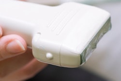
The device, called Scolioscan Air, was developed by a team led by Dr. Zheng Yong-Ping at the university's department of biomedical engineering. It consists of a wireless ultrasound probe with an optical marker mounted at the bottom, a depth camera, and a laptop or tablet computer with dedicated software.
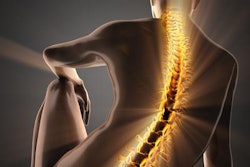
Some 3% to 5% of adolescents in Hong Kong suffer from scoliosis, Yong-Ping and colleagues noted, and its incidence is increasing. That's why early detection of the condition is so important, according to the researchers.
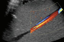
"At present, x-ray imaging is the clinical gold standard for scoliosis assessment, but radiation exposure may pose increased risk for cancer," the team wrote. "Clinical trials have proven the novel technology is very reliable, with an accuracy of curve measurement comparable to x-ray assessment. Moreover, it can obtain images in any posture, provide vertebra rotation and muscle-related information, and form a 3D spinal model for the three-dimensional analysis of deformity of spine," -- achievements that are not possible with common x-ray systems, the researchers noted.
The portability of the device could make scoliosis screening even more accessible, Yong-Ping and colleagues said in a statement released by the university.
"With this innovation, we can now literally bring the device and mass screening service to the youngsters anywhere, anytime," the group wrote. "It would facilitate the implementation of school-based scoliosis screening to detect and treat spinal curvatures before they become severe enough to cause chronic pain or other health issues among adolescents."