Ultrasound users have had a clear message for the imaging industry: Make the modality even easier to use and develop tools that optimize particular applications.
The companies have listened, and at this year's ECR, they are presenting new or upgraded devices that address these demands with software geared toward specific applications such as carotid, liver, obstetrics and gynecology (ob/gyn), and vascular imaging, as well as tools that help users to get the best image during the first scan, improve contrast ultrasound, and fuse ultrasound images with those from other modalities.
Canon Medical Systems is showing advances in its portfolio, including the Aplio i-series, Aplio a-series, Xario g-series, and the Viamo point-of-care systems.
The company's Aplio i800 now features a 33-MHz linear-array transducer that is designed to offer better spatial resolution below 50 microns and improved resolution and detail for B-mode and color flow imaging. Its Smart Sensor 3D allows users to take 3D volumes with a standard linear or convex transducer. The system is particularly suitable for superficial subcutaneous imaging and examinations of small joints and superficial nerves, as well as for superficial vascular evaluations.
 Image of Achilles tendon. Coronal view acquired on the Aplio i800 system with Smart 3D at 24 MHz. Image courtesy of Canon.
Canon is also showcasing its Xario G series, Xario 200G and Xario 100G, which offer up to eight hours of battery-powered, cordless function, allowing the equipment to be used across a variety of locations in a hospital or clinic.
In its booth, GE Healthcare is exhibiting its Logiq E10 ultrasound scanner, which incorporates cSound Architecture using artificial intelligence technology similar to that used to power driverless cars and for 3D video gaming. The scanner acquires and reconstructs data much like MR and CT systems do, and it eliminates the need for focal zones. It also includes cloud connectivity and advanced reconstruction algorithms.
The vendor is also promoting those Logiq E10 features that address the ergonomic challenges ultrasound users face due to repetitive examination steps, one of which is Automated Lesion Segmentation, powered by Edison, which eliminates the need to measure lesions manually by providing a trace of the lesion and corresponding area.
Philips Healthcare is launching a new version of the Epiq system, Epiq Elite, with two software packages, one for vascular imaging and another for ob/gyn applications. Epiq Elite includes an HD display for improved off-angle viewing; on the vascular imaging side, it features the xMatrix linear transducer as well as an upgraded 2D transducer; for ob/gyn applications, it features the V9-2 high-frequency pure wave transducer that allows clinicians to take detailed images as early as possible in pregnancy.
"Clinicians are under continued pressure to image more patients faster, and to be able to accurately reproduce exam results," said Jeff Cohen, vice president of general imaging and women's healthcare ultrasound. "The tools we have developed for Epiq Elite address those challenges."
Siemens Healthineers is highlighting the Acuson Sequoia and Juniper scanners. Acuson Sequoia features a deep abdominal transducer, a 1.2-3.5-MHz ultrasound probe that produces penetration of up to 40 cm. It also boasts the company's BioAcoustic technology, which improves contrast-enhanced ultrasound (CEUS) bubble longevity, allowing for longer view time of contrast agents.
Acuson Juniper's small footprint allows it to be used in a variety of environments. It features 16 transducers and an LED monitor and touch panel, as well as a protective sheet for its keypad to facilitate its use in sterile environments.
Esaote is promoting a portable ultrasound series called MyLab X. It includes three devices: MyLab X5, MyLab X6, and MyLab X7. The series covers applications ranging from abdominal, endocrinological, and cardiovascular to women's health and general imaging, and each offers an increasing mix of features: MyLab X5's tools include stress echo and needle visibility, while MyLab X6 uses ElaXto for measuring tissue elasticity and XLight for volumetric rendering, and MyLab X7 adds Q-Pack for multimodality quantification of curve analysis of contrast perfusion, scope for low/high-frequency modulation, microV for hemodynamic evaluation, and contrast tuned imaging for improving the diagnostic performance.
The company is showcasing two more highly portable devices: MyLab Omega, a general imaging scanner that features XView+, a tool for reducing speckle artifacts, and MyLab Sigma, a laptop-sized scanner designed for ob/gyn applications.
Hitachi is exhibiting three ultrasound scanners: Arietta 850 SE, Arietta 65, and Arietta 50. Arietta 850 SE is an upgraded version of Arietta 850 and features a 23-inch LCD monitor, shear-wave measurement and real-time tissue elastography for the assessment of liver stiffness, and Real-time Virtual Sonography, which offers image fusion for interventional procedures. Arietta 65 features Smooth Workflow and Superb Imaging tools and is useful for real-time elastography and auto intima-media thickness (IMT) measurement, while Arietta 50, the most compact model of the series, features a 21.5-inch monitor and can be used across various applications.
 Color flow cardiac image acquired on Arietta 65 system. Image courtesy of Hitachi.
The company is also demonstrating Futus, another compact system that supports auto IMT and elastography, as well as ob/gyn applications.
Hologic is featuring its Viera portable ultrasound system, a wireless, handheld device. The scanner delivers high-resolution images directly to a smart device from the point of care, and can transmit images to smart devices and PACS networks in offices, examination rooms, and surgical suites.
Visitors to Mindray Medical's booth can learn about upgrades to the Resona 7 ultrasound platform, which is based on the company's Zone Sonography technology and features a range of upgrades that include an expanded transducer selection and improvements such as Sound Touch shear-wave elastography, iFusion with Respiration Compensation, and V-Flow and radiofrequency data-based quantitative analysis on vessel stiffness.
Sound Touch uses Ultra Wide Beam Tracking technology, which produces quantification metrics and visual displays of tissue stiffness. Ultra-Wideband Non-Linear (UWN+) contrast-enhanced imaging uses harmonic and fundamental signals to boost contrast and temporal resolution, while its iFusion tool supports the synthesis of routine B-mode images as well as fusion in CEUS and color/power modes. Mindray's V-flow displays hemodynamic information via speckle-tracking data taken from three plane waves.
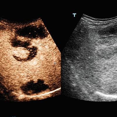
 Contrast-enhanced ultrasound scan of liver lymphoma, using Ultra-Wideband Non-Linear (UWN+) technology. Image courtesy of Mindray.
Samsung is highlighting a software package, SonoSync. The software provides network service to transmit real-time ultrasound examinations so that clinicians at different locations can view images simultaneously. SonoSync can be connected to smartphones, tablets, laptops, and desktop PCs.
The company is also introducing HERA (hyperaperture enhanced reconstruction architecture) W10, which features the Crystal Architecture tool. Crystal Architecture includes Samsung's CrystalBeam beamforming technology and CrystalLive processing engine, and features its S-Vue transducer. HERA W10 also includes ShadowHDR, which creates a shadow-suppressed image; MV-Flow, an alternative to color Doppler for visualizing slow-flow microvascularized structures; and LumiFlow, which offers 3D visualization blood flow images.
SuperSonic Imagine is promoting Aixplorer Mach 30, which includes the company's UltraFast imaging technique to optimize conventional B-mode imaging, as well as its SWE Plus shear-wave elastography software, which helps clinicians visualize anatomical images in real-time while also superimposing color mapping of tissue stiffness. The scanner features SonicPad, a touch pad designed to reduce user fatigue, as well as a variety of transducers, including a high-frequency abdominal probe.
Originally published in ECR Today on 3 March 2019.
Copyright © 2019 European Society of Radiology