Black bone MRI may be a viable, radiation-free alternative to CT for the evaluation of children presenting with abusive head trauma -- particularly for detecting skull fractures, according to a Friday presentation at RSNA 2018 in Chicago.
The researchers, led by presenter Dr. Stephen Kralik of Texas Children's Hospital, examined the potential value of black bone MRI -- a technique that enhances the contrast of bone on MRI scans -- for assessing acute pediatric head trauma. They found that radiologists were able to detect skull fractures nearly as accurately on 3D reconstructions of these MRI scans as they were able to on 3D reconstructions of CT scans.
"Black bone imaging with 3D reconstruction is a promising tool to detect calvarial fractures in young children with suspected abusive head trauma," Kralik told session attendees.
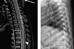
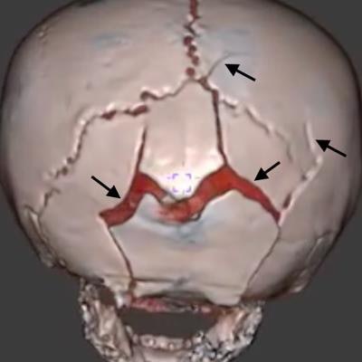
 An 18-month-old girl with multiple skull fractures (arrows) visible on 3D head CT and 3D black bone MRI scans. Image courtesy of Dr. Stephen Kralik.
Black bone MRI
The current gold standard for the initial imaging of suspected abusive head trauma is a CT exam, he noted. CT offers a high sensitivity for detecting acute bleeds and fractures, as well as a high level of accessibility from the emergency department. However, there is growing concern over the potential harms associated radiation exposure from head CT, especially for younger patients.
MRI, like CT, has a high sensitivity and specificity for intracranial traumatic findings, and it has proved useful in the identification of craniofacial pathologies in prior research, without exposing individuals to radiation. Yet its use is reserved primarily for detecting soft-tissue injuries, such as microhemorrhages, and not bone injuries.
Coined in 2012 by Eley et al, "black bone" MRI refers to the acquisition of MRI scans using a short repetition time/echo time (TR/TE) and a low flip-angle, usually with pointwise encoding time reduction with radial acquisition (PETRA). This MRI sequence results in high image contrast between bone and soft tissues and only requires four minutes of imaging time.
"A demonstration of black bone imaging that is comparable to head CT for fracture detection would have value in allowing reduction in ionizing radiation and reduce the need for head CT in trauma patients," Kralik said.
Seeking to determine the effectiveness of black bone MRI in detecting skull fractures, Kralik and colleagues acquired the CT and black bone MRI scans of 34 pediatric patients who presented to the emergency department with potential abusive head trauma. The patients' median age was 4 months.
The researchers then created 3D volume-rendered images of the CT and black bone MRI scans using computer software. Two neuroradiologists independently reviewed these images for the presence, location, and number of skull fractures, with any discrepancies resolved by a third neuroradiologist.
'A promising tool'
Among the pediatric cases examined, there was an 18% incidence of skull fracture based on head CT exams. This comprised a total of 20 distinct fractures, all of which occurred in the parietal or occipital bones.
The radiologists detected 95%, or 19 out of the 20 skull fractures, on black bone MRI, with a sensitivity of 83% and a specificity of 100%, compared with the gold standard of CT. No black bone imaging datasets required repeat imaging. In addition, CT and black bone MRI had an interreader concordance of 0.89 for fracture diagnosis -- indicating almost perfect agreement for the modalities.
This clinical feasibility study demonstrated considerably higher accuracy for black bone MRI, compared with previous research by Dremen et al, who reported a sensitivity of 67% and specificity of 88%. The use of 3D reconstructions in the current study may have led to the improved fracture detection, Kralik noted.
Though the difference was not statistically significant, the sensitivity of black bone MRI was considerably lower when performed with 3-tesla MRI instead of 1.5-tesla MRI, and on patients who were not sedated.
This discrepancy may be attributed to motion artifact as well as the young age of the patient population, he noted. Another limitation of the black bone imaging technique may be its inadequacy in revealing fractures at air-bone interfaces of the skull, such as the mastoid portion of the temporal bone.
"Further refinement in the technique and readily available 3D volume rendering could allow and facilitate its incorporation into the standard of care," he said. "Larger studies are needed to confirm these findings for potential black bone MRI to replace head CT in the neuroimaging evaluation of patients with potential abusive head trauma."