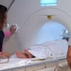
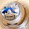
Among the many challenges facing children with cystic fibrosis is them having to hold their breath during imaging. With that in mind, the researchers sought to determine if respiratory gating with a stationary digital chest tomosynthesis system, featuring the carbon nanotube x-ray source, can improve the scanning experience.
Dr. Elias Taylor Gunnell, currently with Memorial Hospital in Savannah, GA, and colleagues recruited 13 pediatric cystic fibrosis patients (average age, 9.6 ± 3.4 years) who were scheduled to undergo chest radiographs. After their chest x-rays, the patients underwent respiratory-gated stationary digital chest tomosynthesis with the carbon nanotube x-ray technology.
Three pediatric radiologists were tasked with comparing the chest radiographs with the stationary digital chest tomosynthesis results and also assessing motion blur and cystic fibrosis pathology. In addition, the researchers performed a retrospective analysis to create a second set of tomosynthesis images, which corrected for first-time images with some blurring from patient motion.
Among the findings, the readers observed larger pixel widths of the diaphragm border with the original digital chest tomosynthesis images, compared with the chest x-rays. In addition, image blurring could be significantly decreased with the corrected digital chest tomosynthesis images.
Respiratory-gated digital chest tomosynthesis is quite valuable using the carbon nanotube x-ray source and would allow for significantly reduced respiratory motion blur and more productive scans for the young patients, Gunnell and colleagues concluded.