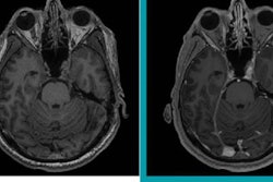
Swiss researchers have confirmed the presence of gadolinium in cerebrospinal fluid (CSF) within a few hours of an MRI scan with a macrocyclic gadolinium-based contrast agent (GBCA). The findings indicate that CSF could be the pathway through which gadolinium enters the brain, they concluded in the September issue of Radiology.
Researchers have been trying to determine the mechanism by which gadolinium is retained in brain tissue -- in some cases years after contrast administration. Recent studies have examined cerebrospinal fluid as the possible mechanism by which gadolinium passes from the bloodstream and into brain tissue.
For the current study, Dr. Florian Berger from the University of Zurich and colleagues retrospectively analyzed 60 CSF samples from 57 patients (median age, 50 years; range, 3-92 years). All subjects had received one MRI scan with gadoterate meglumine (Dotarem, Guerbet) within 60 days of CSF being taken between January and December 2016 (Radiology, September 2018, Vol. 288:3, pp. 703-709).
The researchers divided the samples into three groups based on the time between GBCA administration and CSF extraction. Group 1 consisted of 31 samples acquired between zero and eight hours after MRI, group 2 had 13 samples acquired eight to 48 hours after MRI, and group 3 had 16 samples acquired two to 60 days after MRI.
In addition, 24 subjects (median age, 60.5 years; range, 19-79 years) served as a control group. Of this group, 22 had not received contrast with MRI, while the other two had GBCA-enhanced MRI scans one and three years, respectively, before CSF samples were taken.
The researchers found gadolinium in CSF samples from all 57 patients, with an overall mean concentration of 816.1 ± 746.6 ng/mL (range, 0.1-3,481.9 ng/mL). Mean CSF gadolinium concentrations were highest in the first eight hours after GBCA administration and decreased thereafter.
| CSF gadolinium concentrations after MRI scan based on time | |||||
| Group 1 | Group 2 | Group 3 | |||
| Mean concentration (ng/mL) | 1,151.5 ± 734.6 | 871.7 ± 586.1 | 121 ± 296.3 | ||
| Median time between GBCA administration and CSF sampling | 4.6 hours | 23.4 hours | 5.7 days | ||
CSF gadolinium levels were greatest among three patients with chronic renal insufficiency (2,257, 2,140, and 532 ng/mL at 5.5 hours, 25.5 hours, and three days after MRI, respectively), indicating a "prolonged GBCA elimination from CSF," Berger and colleagues wrote.
In other findings, the authors noted that gadolinium was detected in individuals who presumably had an intact blood-brain barrier. They also noted that gadoterate meglumine was cleared almost completely from the CSF within 48 hours after contrast administration.


