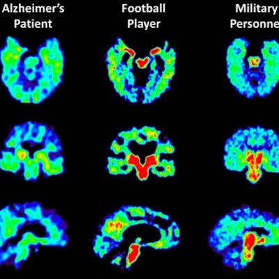
Researchers have found that changes in PET scans of military personnel with mild traumatic brain injury (TBI) are similar to those seen in retired football players with suspected chronic traumatic encephalopathy (CTE), according to a study published online July 17 in the Journal of Alzheimer's Disease.
The changes were seen through the use of the tracer FDDNP, which binds to tau and amyloid on PET. Both proteins have been linked to the onset of mild cognitive impairment, Alzheimer's disease, and dementia.
"This first study of in vivo tau and amyloid brain signals in military personnel with histories of mild TBI shows binding patterns similar to those of retired football players and distinct from the binding patterns in Alzheimer's and normal aging, suggesting the potential value of FDDNP-PET for early detection and treatment monitoring in varied at-risk populations," wrote senior author Dr. Gary Small from the University of California, Los Angeles (UCLA) and colleagues.
Chronic traumatic encephalopathy occurs in people who have had multiple head injuries. The condition is associated with memory loss, confusion, progressive dementia, depression, suicidal behavior, personality changes, and abnormal gait and tremor.
Small and colleagues have been developing FDDNP-PET for the past several years to analyze unusual brain patterns in former athletes with repeated brain injuries. The findings in this study build upon previous research in which UCLA investigators used FDDNP-PET to determine imaging patterns in 14 former professional football players.
For the current study, Small and colleagues performed FDDNP-PET brain scans on seven military personnel, 15 retired players with mild TBI histories and cognitive and/or mood symptoms, 24 people with Alzheimer's disease, and 28 cognitively normal control subjects.
The researchers found significantly greater binding of FDDNP in military personnel than in the controls in the amygdala, midbrain, medial thalamus, pons, frontal, and anterior and posterior cingulate regions (p < 0.01-0.0001).
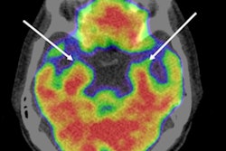
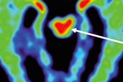
 Warmer colors indicate a higher concentration of the tracer FDDNP, which binds to abnormal proteins in the brain. Image courtesy of UCLA Health.
The FDDNP binding patterns were similar between the military and former football players -- except in the amygdala and striatum (p = 0.02-0.003). Compared with the Alzheimer's group, military personnel had significantly greater binding in the midbrain (p = 0.0008) and pons (p = 0.002) but significantly lower binding in the medial temporal, lateral temporal, and parietal regions (all p = 0.02).
"Larger studies using FDDNP and brain scans in people at risk for chronic traumatic encephalopathy are needed to confirm the usefulness of FDDNP as a diagnostic tool," concluded a release from the university. "The ability to diagnose chronic traumatic encephalopathy in living individuals would help scientists observe the progression of the disease, develop treatments, and quantify the scope of CTE among different populations, such as military veterans."