Using MRI scans to view brain lesions in patients with multiple sclerosis (MS), researchers at the University at Buffalo (UB) are contradicting a decades-long belief on how to determine whether the condition will progress. Their results were published online June 1 in the Journal of Neuroimaging.
The five-year study suggests that the atrophy of brain lesions is a key indicator of disease progression. Until now, the appearance of new or expanding brain lesions was considered an indication that a patient's MS was worsening.
"Using the appearance of new brain lesions and the enlargement of existing ones as the indicator of disease progression, there was no sign of who would develop disability during five or 10 years of follow-up," said senior author Dr. Robert Zivadinov, PhD, director of the Buffalo Neuroimaging Analysis Center. "But when we used the amount of brain lesion volume that had atrophied, we could predict within the first six months who would develop disability progression over long-term follow-up."
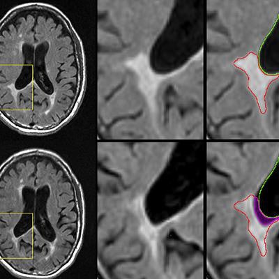
 MR images represent an atrophied, or disintegrating, lesion on baseline (top row) and follow-up scans (bottom row). Part of the original lesion (bottom right, magenta region) has disintegrated into cerebrospinal fluid over the intervening period. Nearly 20% percent of the lesion was lost over time. Images courtesy of Michael Dwyer, PhD.
Currently, MS patients undergo MRI scans so clinicians can see whether new lesions are forming or existing lesions are growing. If lesions disappear, it has been considered a sign of improvement.
However, the researchers found no correlation between the development of more or larger lesions and the worsening of multiple sclerosis.
The five-year study included 192 patients with one of three subtypes of MS:
- 18 people had clinically isolated syndrome, the earliest stage of the disease.
- 126 people had relapsing-remitting MS, an intermediate stage.
- 48 people had progressive MS, which is the most severe stage.
Three-tesla MRI scans were used to measure lesion volume at the start of the study and through annual follow-up MR imaging for more than five years. The scans revealed that atrophied lesion volume was the only factor that correlated significantly with a change in patient disability (r = 0.192 relapsing MS, r = 0.317 progressive MS, p < 0.05).
Another important finding is that atrophied brain lesions were a more robust predictor of MS progression than whole-brain atrophy, which has been the most accepted biomarker of MS-prompted neurodegeneration.
"Our data suggest that atrophied lesions are not a small, secondary phenomenon in MS, and instead indicate that they may play an increasingly important role in predicting who will develop a more severe and progressive disease," Zivadinov concluded in a release from the university.