Chinese researchers found that digital tomosynthesis with a metal artifact reduction algorithm was better than either CT or conventional radiography for assessing the integration of hip implants with bone. Their findings were published in the May issue of the Journal of Arthroplasty.
Digital tomosynthesis with metal artifact reduction (TMAR) had a sensitivity of nearly 74% for detecting the cementless osteointegration of hip implants with bone, compared with 50% for radiography and 36% for CT. The specificity of TMAR also exceeded the performance of the other modalities, according to a group led by Dr. Shengjie Guo (J Arthroplasty, Vol. 33:5, pp. 1579-1587).
"Our study revealed that the sensitivity, specificity, positive predictive value, and negative predictive values for TMAR for assessing osteointegration in cementless hip arthroplasty were significantly improved ... compared with those for radiography and conventional CT," Guo and colleagues wrote.
Get the lead out
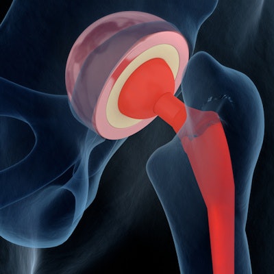
The use of cementless techniques to implant hip prostheses has become increasingly popular, and the number of these types of procedures is expected to rise in the coming years. But a major issue is checking the stability of implants to make sure biologic fixation between the implant and bone is occurring.
Imaging techniques like CT and radiography have been used for this purpose, with the "spot-weld" sign a classic indication that biologic fixation is occurring around the implants. But the effectiveness of radiography can be obscured because structures can overlap each other and there is less spatial resolution regarding depth. Meanwhile, the metal in the implant can cause artifacts on CT scans, making it harder to visualize spot-weld signs.
Could digital tomosynthesis help? The scanning tube head of a tomosynthesis system acquires multiple images that are reconstructed into a single volume, giving a depth of resolution that radiography lacks. It also avoids the metal artifact problem common with CT while delivering a lower radiation dose to patients.
Guo and colleagues tested the use of their digital tomosynthesis system (Sonialvision Safire II, Shimadzu Medical Systems) with a metal reduction algorithm in a group of 24 patients, with a total of 13 femoral stems and 14 acetabular cups implanted using the cementless technique. All had solid evidence of biologic fixation, and patients received three imaging exams: conventional radiography, CT, and tomosynthesis with TMAR.
The researchers then rated the ability of the three techniques to detect the spot welds that were telltale signs of osteointegration. Images were evaluated by a group of seven orthopedic surgeons; implants retrieved from the patients were used as the reference standard.
TMAR performed better than either CT or conventional x-ray, the researchers found. All results were statistically significant (p < 0.017).
| Use of TMAR for detecting osteointegration, by implant type | |||
| CT | X-ray | TMAR | |
| Femoral stem | |||
| Sensitivity | 36.4% | 50.4% | 73.8% |
| Specificity | 90.9% | 87.8% | 94.3% |
| PPV | 59.5% | 60.3% | 82.7% |
| NPV | 79.5% | 82.8% | 90.7% |
| Acetabular cup | |||
| Sensitivity | 45.1% | 45.9% | 60.2% |
| Specificity | 73.5% | 66.4% | 86.4% |
| PPV | 61.2% | 56.5% | 80.8% |
| NPV | 59.0% | 56.4% | 69.5% |
Detecting osteointegration means that forces are being transmitted "in a stable biomechanical pattern between prostheses and bone," which is the key sign on imaging that biologic fixation is occurring, the group wrote. TMAR offers a new tool for detecting this process, according to Guo and colleagues.
"The use of TMAR leads to greater contrast and removal of metal artifacts in the in-focus plane," they wrote.
They noted that there were some drawbacks with TMAR compared to the other modalities. CT can be reconstructed in any plane down to a slice thickness of 0.5 mm, while TMAR images require acquisitions in both coronal and sagittal views at a minimum thickness of 2 mm. This can give CT more flexibility in identifying spot welds.
However, TMAR has the advantage of a lower intrinsic radiation dose than CT; what's more, the technique's radiation can be limited to a small area, further reducing dose.
In the end, tomosynthesis with metal artifact reduction can be a useful tool for making sure hip implants are successfully integrating with bone.
"Our findings indicate that in comparison with radiography and conventional CT, TMAR can greatly improve the accuracy of osteointegration detection in cementless hip arthroplasty components," they concluded.