RSNA 2017 is almost here, and considering all the developments in advanced visualization that have taken place this past year, the timing could not be better.
3D printing has taken great strides, and numerous workshops, courses, and presentations in McCormick Place will cover the basics of 3D printing -- particularly how to use it and make it cost-effective -- as well as the flashier aspects of incorporating 3D printing into medical practice.
Collectively, this year's roundup of 3D printing sessions promotes the technology's role in bridging the gap between a theoretical understanding of procedures and actually performing them. Researchers will describe how they used 3D-printed models to simulate arthrography, angiography, valve replacement, and CT- and MR-guided thermal ablation. They will also expound how 3D printing can buttress the development of implants and what type of printing materials are best-suited for visualization.
Also stepping into the spotlight this year is 4D imaging. Investigators have been using 4D CT to catch invasive or growing pathology in specific organs, whereas they have reserved 4D MRI more for displaying blood flow and joints in motion. What better way is there to understand anatomical function and assess pathology than to visualize it all in real-time?
Advanced visualization is bringing virtual reality into the front lines as well. Most commonly associated with video games, virtual reality has recently been lending a hand to clinicians, including radiologists.
One research group will showcase a virtual and augmented reality educational platform that allows radiologists to study common cases online and on the go. Another team will explain how it used a virtual reality headset to train radiologists for emergency contrast situations, such as patients having allergic reactions. Extending its reach, virtual imaging can also bolster diagnosis in the brain through real-time virtual sonography, as well as help physicians optimize and individually tailor surgeries, according to presenters.
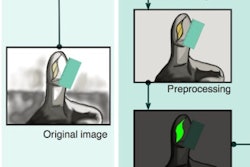
Interest in the latest imaging modalities continues to grow by leaps and bounds. A prime example is the 3D visualization of medical images, which when performed using 3D fusion imaging software, allows for joint assessment of the coronary arteries by combining coronary CT angiography and CT myocardial perfusion images. The computer animation technique of cinematic rendering adds yet another (realistic) dimension to 3D imaging.
Last but not least, attendees interested in understanding how human perception affects image analysis should stop by the Perception Lab in the Lakeside Learning Center to take part in brief, snazzy visual experiments designed specifically for radiologists. Jeremy Wolfe, PhD, from Brigham and Women's Hospital, and Todd Horowitz, PhD, from the National Cancer Institute, will lead the exhibit.
More information on these and similar advanced visualization sessions awaits below. The complete listing of abstracts for RSNA 2017 is available here.