MRI scans of astronauts who spent time at the International Space Station confirm that increased orbital and ventricular cerebrospinal fluids cause vision problems that worsen with extended time in space and linger after the travelers have returned to Earth.
Researchers from the University of Miami observed the link between space travel and a condition known as visual impairment and intracranial pressure (VIIP) syndrome, in which the globe of the eye flattens and the optic nerve can protrude. While the findings signal vision risks for astronauts, there could be residual benefits for earthbound patients with certain eye disorders.
"Our quantitative assessment shows that [VIIP] does not occur in short durations, but only in long duration, and the effect of how long [astronauts] stay in space is critical here," said lead author Noam Alperin, PhD, a professor of radiology and biomedical engineering at the University of Miami. "The methodology that we have developed has been applied to idiopathic intracranial hypertension. It is very likely that it will be applicable for glaucoma."
 Noam Alperin, PhD, from the University of Miami.
NASA's knowledge
NASA has been aware of health risks related to vision in astronauts since 2005. That's when approximately two-thirds of U.S. astronauts who spent time on the International Space Station began showing symptoms of VIIP and changes in ocular structure. The agency already knew that weightlessness during space flights could adversely affect the cardiovascular system, so naturally its focus was to explore that cause for the eye problems.
As part of NASA's investigation of VIIP syndrome, it contacted Alperin and colleagues, who are well-known for their research on ocular and vision issues related to cerebrospinal fluid biomechanics in the brain.
"NASA called us. They told us, 'Miami, this is Houston. We have a problem,' " Alperin quipped to AuntMinnie.com. "So we agreed to use our MRI protocol and perform scans before and after space flights with a long duration and a short duration."
To begin, the researchers separated astronauts into two groups to better determine how time spent in the Space Station affects vision. The long-duration group consisted of seven astronauts (mean age, 46.1 years ± 5.5 years) who spent a mean of 118 days (± 22 days) in space. The short-duration group consisted of nine astronauts (mean age, 46.9 years ± 2.6 years) who spent a mean of only 14.1 days (± 1.6 days) in space (Radiology, April 21, 2017).
All 16 astronauts had MRI scans of their brains and the orbits of their eyes on a 3-tesla scanner (Magnetom Verio, Siemens Healthineers) between 2010 and 2014 before they went into space and when they returned.
"MRI provides reliable geometric details of the eye globe and the orbital cerebrospinal fluid spaces and, at the same time, provides us with information about the changes that occurred in the brain," Alperin explained. "That gave us the ability to find a correlation between changes in the ocular structure and changes in the brain and understand the association between the two."
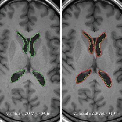
An automated quantitation algorithm was then used to measure pre- to postflight changes in orbital and ventricular cerebrospinal fluid (CSF) volumes and in brain tissue volumes. The algorithm is designed to identify the center of the eye globe and lens as landmarks of the optical axis, along with the distal end of the optic nerve and the surrounding CSF region.
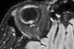
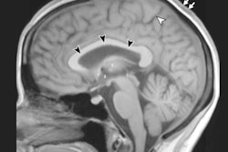
 MR images show the orbit and brain with the automated delineation of orbital and ventricular CSF spaces obtained before flight (green) and after flight (red) for a long-duration male astronaut. Orthogonal planes of the orbital CSF demonstrate postflight expansion and elongation of the CSF space around and along the length of the optic nerve. Images courtesy of Radiology.
The eyes have it
In analyzing the pre- and postflight MRI scans and quantitation results, the researchers found that globe flattening of the astronauts' eyes increased significantly in the long-duration cohort (0.031 ± 0.019), compared with results from astronauts who spent less time in space (0.001 ± 0.006) (p < 0.00002).
The before and after nerve protrusion results also showed significant changes in the long-duration group (0.025 ± 0.013) versus the short-duration group (0.001 ± 0.006) (p < 0.0001).
Those increases in globe flattening and nerve protrusion were associated with significant increases in orbital and ventricular cerebrospinal fluid volumes. Interestingly, brain tissue volumes did not change based on the length of time in space or correlate with globe flattening and optic nerve protrusion.
| Pre- to postflight changes | |||
| Change in orbital CSF volume (µL) | Change in ventricular CSF volume (mL) | Change in white-matter volume (mL) | |
| Short duration | 1.8 | 0 | 3.9 |
| Long duration | 51.0 | 2.9 | 1.5 |
| p-value | 0.0005* | 0.048* | 0.58 |
Controlled environment
"The fact that the astronauts were in the Space Station provided us with the control to understand what changes occur due to the length of exposure to microgravity and remove the short duration when [astronauts] would go for two weeks and come back," Alperin said. "This is a control case where we know what kind of change is associated with long duration."
Of particular concern to NASA is that the vision problems linger with astronauts long after they have come home. Needless to say, the agency is looking for ways to rectify that issue. Alperin, for one, is optimistic.
"I think this problem is very solvable, and the methodologies that we can apply once we understand the problem can account for that," he added. "We have to find the optimal protocol that would enable us to prevent this from developing to the degree that will be damaging."