MRI scans have revealed significant reductions of gray matter in several brain regions of children with moderate or severe obstructive sleep apnea, compared with children who sleep normally, according to a paper published online March 17 in Scientific Reports, one of the Nature journals.
The noted brain regions are associated with movement, memory, emotions, speech, perception, decision-making, and self-control, and the results indicate a loss of neurons or delayed neuronal growth in the developing brain in connection with sleep apnea.
Researchers from the University of Chicago recruited 16 children ages 7 to 11 years with obstructive sleep apnea who underwent neurocognitive testing and MRI brain scans. The MR images and test scores were then compared with results from nine age- and gender-matched healthy children who did not have apnea. The researchers also compared the children with obstructive sleep apnea to 191 children who were part of an existing pediatric MRI database from the U.S. National Institutes of Health.
The MRI scans showed reductions in the volume of gray matter in multiple regions of the brain in children with obstructive sleep apnea. More specifically, the losses were discovered in the frontal cortices, which handle movement, problem-solving, memory, language, judgment, and impulse control; the prefrontal cortices, which are associated with complex behaviors, planning, and personality; the parietal cortices, which are linked to sensory input; the temporal lobe, which is involved in hearing and selective listening; and the brainstem, which controls cardiovascular and respiratory functions.
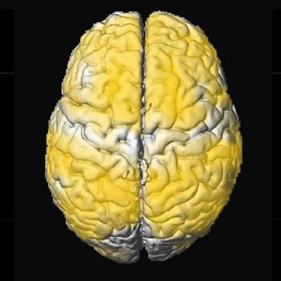
 Images show regions of reduced gray-matter volume (yellow) in subjects with obstructive sleep apnea versus control subjects. Courtesy of Dr. Mona Philby et al.
While the MRI scans illustrate the difference in gray-matter volume from obstructive sleep apnea, the images do not show whether brain cells have shrunk or been lost completely, or exactly when the damage occurred, said study co-author Dr. David Gozal, a professor of pediatrics, in a statement.
Future collaborative studies are planned between researchers at the University of Chicago and the University of California, Los Angeles to attempt to answer such questions.