"In spite of its unparalleled 4D nature, cine functional assessment by cardiac CT is not routinely performed in the current clinical practice due to high radiation," wrote Dr. Kakuya Kitagawa, PhD, in an email to AuntMinnie.com. "But what if it can be performed with less than 1 mSv of radiation right after coronary CT angiography without additional contrast material?
4D functional cine cardiac CT is a technique worth revisiting, considering the low-dose imaging capabilities of the latest dual-source scanners and advanced postprocessing technology, said Kitagawa, who is an associate professor of radiology at Mie University in Tsu, Japan.
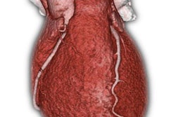
Conventional retrospectively gated 4D volume cine cardiac CT is acquired with ECG-gated multislice scanning with 3D reconstruction of the heart volume, but its clinical use is limited due to radiation that routinely tops 20 mSv. This study relied on the recirculation of contrast media used for coronary CT angiography to acquire the images without additional contrast injection, the investigators wrote in their abstract.
The technique was applied to 10 patients referred for cine cardiac CT before transcatheter aortic valve implantation (TAVI) planning. Retrospective ECG-gated cine CT was acquired at 120 kV using a third-generation dual-source CT scanner. The 20-second scan delay was set to allow the low-dose cine to capture the recirculation of contrast medium injected for a standard CT angiography scan, with 20 axial image series being reconstructed every 5% of the RR interval. Low-dose images were then processed with a nonrigid registration-based algorithm.
For a mean radiation dose of less than 1 mSv, all measurements including end-systolic volume, end-diastolic volume, ejection fraction, and left-ventricular mass by low-dose CT agreed well with standard scans, Kitagawa reported.