It's time for coronary CT angiography (CCTA) to replace conventional invasive angiography for evaluating coronary ischemia in low- to moderate-risk patients, according to the results of a direct comparison of the two techniques published this month in JACC: Cardiovascular Imaging.
In what lead author Dr. Matthew Budoff and colleagues are calling the first head-to-head test of the two procedures, more than 200 patients from five countries underwent both CCTA and quantitative coronary angiography (QCA) along with wire-based fractional flow reserve (FFR) as the gold standard exam.
The researchers found the same moderately good sensitivity, specificity, accuracy, and positive and negative predictive values for the two exams. However, CCTA had important additional benefits, such as noninvasiveness and the ability to add FFR-CT for further validation (JACC: Cardiovasc Imaging, February 17, 2016).
"This study shows that invasive angiography and cardiac [CT] are similar in their ability to detect functionally significant stenosis as defined by fractional flow reserve," Budoff wrote in an email to AuntMinnie.com. "Thus, the use of CCTA can replace invasive angiography for diagnosis, without losing accuracy for the clinician who wants to identify flow-limiting lesions for revascularization."
Budoff is a professor of medicine at the University of California, Los Angeles (UCLA) and chair of preventive cardiology at the Los Angeles Biomedical Research Institute in Torrance, CA.
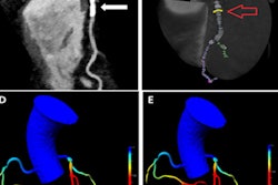
Measuring stenosis
CCTA has long been shown to be a highly diagnostic test that leads to better outcomes than functional testing, but it is always being compared with invasive angiography as a reference standard. Angiography, however, has well-known shortcomings, including a persistent underestimation of coronary plaque burden compared to intravascular ultrasound or optical coherence tomography, particularly in the setting of vascular remodeling. The validation of FFR offers a way to finally compare the accuracy of CCTA and angiography.
Budoff and colleagues compared the diagnostic accuracy of quantitative coronary angiography and CCTA versus invasive FFR measurements. Quantitative angiography and CCTA had not previously battled head to head for the prediction of ischemia as determined by FFR in a large multicenter cohort.
The 17-center multinational study included 252 patients (71% male; mean age, 63 years) who underwent CCTA and invasive coronary angiography with FFR in 407 lesions. All patients had participated in the Determination of Fractional Flow Reserve by Anatomic Computed Tomographic Angiography (DeFACTO) study, the results of which were reported previously. DeFACTO was designed to assess hemodynamically significant coronary disease, defined by invasive FFR, in a population with suspected coronary disease.
CCTA was performed on 64-detector-row CT scanners with either prospective or retrospective electrocardiogram (ECG) gating. The exams were interpreted at the central core laboratory in Torrance.
Angiography exams were interpreted at an independent core laboratory at the University of British Columbia in Vancouver using commercially available software. FFR (PressureWire Certus, St. Jude Medical) was performed at the same time as invasive angiography. Per-patient diagnostic performance comparisons looked at accuracy, sensitivity, specificity, positive predictive value, and negative predictive value.
No difference in accuracy
The study confirmed that measurements derived from quantitative angiography and CCTA perform similarly for the prediction of lesion-specific ischemia. This suggests "that coronary CTA may be used as an alternative to assess luminal stenosis and to serve as gatekeeper to FFR measurements in patients presenting with chest pain syndromes," the authors wrote.
FFR revealed ischemia in 151 (37%) of the 407 lesions; the area under the curve (AUC) for identifying ischemia-causing lesions was similar for angiography and CCTA. In addition, there was no difference between them for determining ischemia within the left anterior descending, left circumflex, and right coronary arteries.
| Per-patient performance of angiography vs. CCTA | ||
| Measure | Quantitative angiography | CCTA |
| Accuracy | 71% | 69% |
| Sensitivity | 74% | 79% |
| Specificity | 70% | 63% |
| Positive predictive value | 59% | 55% |
| Negative predictive value | 82% | 83% |
Quantitative angiography and CCTA each have their strengths and weaknesses, the authors noted. Angiography has higher spatial and temporal resolution than CCTA, which is thought to be less accurate in quantifying the extent of stenosis.
"However, coronary CTA has higher contrast resolution, allowing plaque to be seen, which has been shown to improve prediction of FFR," they wrote. "Furthermore, the [3D] nature of coronary CTA may allow for differential assessment of stenosis severity compared with the [2D angiography]. This approach may answer the question as to why lower anatomical accuracy does not affect the accuracy in assessing homodynamic significance."
Importantly, CCTA also enables the use of an FFR surrogate, FFR-CT, to enhance the diagnostic potential of CCTA without additional scanning or radiation.
Editorial: Replace invasive angiography
In an accompanying editorial, Dr. Armin Arbab-Zadeh, PhD, from Johns Hopkins University wrote that these "results are important because they demonstrate yet again that conventional angiography is not superior to CT scanning for the diagnosis of [coronary artery disease (CAD)] (in this case, hemodynamically significant CAD) compared with an independent reference standard."
The shortcomings of quantitative angiography are well-known; for example, it does not agree well with intravascular ultrasound on the severity of stenosis.
"Indeed, agreement between intravascular ultrasound and CT scanning regarding lumen area is greater than that by intravascular ultrasound and QCA." he wrote. "This should not be surprising because both CT scans and intravascular ultrasound allow comprehensive vascular assessment in contrast to the limited, [2D] projections available by conventional angiography."
Recent studies have shown that the functional significance of lesions should guide patient management. The finding that CCTA and quantitative angiography are equivalent "suggests that due to lower costs and improved safety, coronary CTA may act as a gatekeeper and first-line test to triage patients to medical therapy or invasive evaluation with FFR (with potential revascularization)," he wrote.
While angiography employed its best quantitative methods, Budoff and colleagues relied on a visualization to estimate stenosis, forgoing the many other tools such as lesion length and area stenosis available for stenosis analysis with CCTA.
"Thus, CT scanning sent only its 'B' team and still held its own compared with our current gold standard for the diagnosis of CAD," Arbab-Zadeh wrote.
Given CT's performance, "why is it then that we still puncture arteries and advance catheters in the aortas of >1 million patients every year for the diagnosis of CAD?" he wrote. "We do so simply because we have no data on patient management based on a CT scan-guided approach directly compared with the standard approach of invasive angiography."
Convincing the medical community and patients that CT is equivalent to angiography will require CT providers to demonstrate that management decisions based on CCTA yield the same or better results for patients -- a daunting task that will take years but offers big opportunities, he added.
The current generation of scanners has drastically lowered radiation dose and improved accuracy, and patients already prefer CCTA for coronary disease testing.
"We are almost there," Arbab-Zadeh wrote.