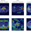
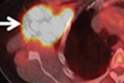
A group led by Dr. Onofrio Catalano of Massachusetts General Hospital imaged 49 consecutive women with newly diagnosed invasive ductal breast carcinoma using the aforementioned modalities, as well as contrast-enhanced PET/CT, prior to treatment.
Whole-body PET/MRI correctly staged all 49 subjects, while PET/CT correctly staged 38 (78%), the researchers found. PET/MRI was also best at detecting malignant lymph nodes, with a detection rate of 96%, followed by whole-body diffusion-weighed imaging at 76%.
The potential benefits for patients are "more accurate staging than achievable with other imaging techniques, and less radiation dose if compared to PET/CT," Catalano wrote in an email to AuntMinnie.com.
"PET/MRI should be made available to stage patients with breast cancers, especially in the case of questionable metastatic disease with other techniques, clinical suspicion of advanced disease that is not confirmed by traditional staging techniques, and initial staging," he added.
Catalano and colleagues hope to recruit more patients to confirm their preliminary results and to refine a protocol that would benefit the largest number of patients with breast cancer.


