Preclinical imaging technology developer MILabs is touting a study published online in Molecular Imaging and Biology that concluded 4D microSPECT works for studying murine cardiac function.
Researchers at Duke University evaluated a new generation microSPECT with resolution less than 0.5 mm as a feasible imaging modality for studying murine cardiac function, comparing it with microCT. The team used MILabs' U-SPECT-II/CT, which includes a 0.35-mm-resolution collimator, and an in-house CT system developed for dynamic imaging applications (Mol Imaging Biol, September 2013).
Dr. Allan Johnson and colleagues found that the two modalities showed excellent correlation in measured cardiac function, despite differences in spatial resolution and contrast-to-noise ratio, according to MILabs.
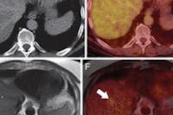
An additional advantage of microSPECT was that quantitative analysis of both cardiac function and multiple myocardial radiotracer distributions could be performed from a single 4D acquisition, eliminating the need for multiple contrast agents or additional acquisition protocols. Also, U-SPECT could accurately identify the regions of ischemia or infarction that were not visible in the microCT images, MILabs said.