The adoption of PACS hasn't eliminated the problem of lost x-ray films, as any healthcare facility knows. But Japanese researchers believe they've found an answer through a computer algorithm that locates misfiled chest radiographs based on unique anatomical data, which they call "biological fingerprints," found in each x-ray.
Even in the digital era, a medical imaging study can still be lost if incorrect patient information is added to the data file that accompanies it. This could cause the exam to be misfiled and stored in the wrong patient's folder, which could lead to radiologists interpreting the wrong images, according to a research team led by Risa Toge of Kyushu University.
Toge's team presented early research on the algorithm at the 2008 American Association of Physicists in Medicine (AAPM) meeting. Since then, they've revised the algorithm to make it even more accurate in finding matches for lost x-rays; they published their findings on June 15 (ahead of print) in Radiological Physics and Technology.
The problem of misfiled x-rays
Toge's team, which included imaging informatics specialist Kunio Doi, PhD, from the University of Chicago, determined in previous research that misfiled x-rays could be a problem: Of 279,222 studies acquired over approximately two years at a university hospital in Japan, 327 studies, or 0.117%, were misfiled and temporarily lost.
"Misfiled images in a PACS environment may create serious medical accidents in hospitals," Toge's team wrote.
Japanese hospitals have developed a "kenzo system" for checking images after they are acquired but before they are stored to confirm that patient information is correct. But such a manual protocol can be time-consuming, and an automated image-searching system for finding misfiled images quickly without requiring personnel time would be useful.
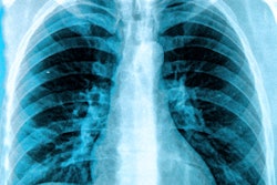
Toge and colleagues developed an automated mathematical algorithm that creates a "biological fingerprint" for an image based on anatomical information in the x-ray. The fingerprints are based on five attributes with distinct anatomic structures, such as cardiac shadow, lung apex, superior mediastinum, right lower lung, and the whole lung field.
The mathematical algorithm examines the biological fingerprint of the misplaced image and searches for a match in the PACS database, using the assumption that two different images of the same patient will be more similar than any images from different patients. By matching the five attributes, it produces a correlation index score, and the image with the highest score is identified as the correct match for the misfiled image.
The researchers tested the algorithm in 200 pairs of chest radiographs, drawn randomly from a database of more than 36,000 chest radiographs of individuals who participated in a lung cancer screening program with computed radiography in Iwate Prefecture.
They also varied the weighting of each of the five attributes in producing the correlation index score, on the assumption that some anatomical features could be more useful than others in matching images. For example, cardiac shadow and right lower lung attributes were more useful than whole lung field and lung apex values.
Using their original set of weighting factors, the researchers found that the algorithm was able to produce a correct match between 156 of 200 pairs of images (78%). They then altered the weighting factors, and this change boosted the match accuracy to 175 of 200 pairs (87.5%). An additional change, applying image rotation to the overlapped region of two images, boosted accuracy to 179 of 200 cases (89.5%).
What about the remaining cases that didn't match? Due to the small number of such cases, these could be managed manually by radiology personnel, Toge and colleagues wrote.
The method requires at least one previous chest radiograph stored in the proper location as a clue to identify the correct patient for a misfiled image, the authors noted. More refined weighting factors could also be developed.
While the algorithm isn't currently being used in the clinical setting, Toge believes that it could be used as a sort of automated warning system for avoiding filing errors in radiographs before they are saved to a PACS. The warning system would alert users before the image is stored if it detects a mismatch.
If the algorithm proves feasible, it could ultimately be expanded to bedside radiography, other parts of x-ray images, and even images in other imaging modalities.