Although high-performance 3D/4D technology has been available in ultrasound for years, it hasn't routinely been used in all clinical practices. That may change in 2013, however, thanks to a number of key trends that are expected to drive widespread adoption.
Until recently, there was low clinical demand for upgraded 3D/4D technology due to a perceived lack of clinical evidence and a paucity of affordable hardware with the required level of performance. Unconvinced of its clinical advantage, clinicians did not demand 3D technology and, consequently, hardware was not upgraded to keep up with changing software.
Because physicians demand high accuracy from ultrasound images, however, 3D/4D ultrasound imaging is now crucial. It decreases the risk of not having acquired the entire anatomy, as could be the case with a 2D examination. With 3D, the whole anatomy can be acquired with one single scan for many clinical applications. As clinical evidence proves the quality of 3D/4D, clinicians will be more likely to adopt this technology.
More clinical evidence
In general, radiology stakeholders are conservative in their actions and require extensive research before they widely accept new practices. For years, 3D/4D technology has undergone clinical testing and has now started to accumulate clinical evidence. The fact that volumetric scanning leaves no information behind will ensure that all information is acquired and processed, ultimately leading to a faster, more accurate diagnosis.
For example, 3D fetal ultrasound examinations have demonstrated a superior visualization of subtle fetal brain defects and other fetal anomalies after 3D image enhancement has been applied to the 3D volume. Specifically, structures in the near field behind the skull bone are better visualized in the fetal brain with 3D, according to Dr. Anders Selbing, PhD.1
The benefits and prerequisites to using volume US for fetal brain studies were also presented by Dr. Boris Tutschek at last year's International Society of Ultrasound in Obstetrics and Gynecology (ISUOG) annual meeting.2 Tutschek concluded that "volume 3D enables ideal examination setting and correlation of every point in the brain in two or three [orthogonal] planes," and "enables tomographic imaging [comparable with MR and CT]."2 Furthermore, 3D acquisition enables 3D image enhancement using neighboring volume characteristics.
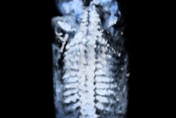
 |
| Fetal brain: Upper row contains nonprocessed 3D-acquired images in A, B, and C planes. Lower row contains enhanced 3D-acquired images in A, B, and C planes. Note the structures in the near field behind the skull bone are becoming visible, leading to new diagnostic possibilities. Images courtesy of Dr. Anders Selbing, PhD.1 |
At the 2012 ISUOG meeting, doctors not only presented on the positive power of 3D, they also used clinical evidence to make their arguments. Specifically, Dr. Beryl Benacerraf of Harvard Medical School showed that a 3D coronal image is a necessary part of a pelvic scan for positioning the arms of an intrauterine device (IUD) and that "with 3D there is a possibility to distinguish a septate from a bicornuate uterus since the three orthogonal planes are necessary to evaluate the shape of the cavity."3 Dr. Valentina Chiappa concluded that "3D-US seems to be an accurate diagnostic tool in assessment of parametrial infiltration, with comparable results to MR in all cases with evident infiltration."4
The real risk
As two dimensions have been successful thus far, radiologists in general have been comfortable with 2D ultrasound, unsure if the unclear benefits of 3D/4D will outweigh the risks of turning to the unknown. However, 3D is today approved to be used in daily practice, and the real concern is that a clinician may risk missing something in a 2D scan. A study by Goldberg, Forsberg, and Lev-Toaff at Thomas Jefferson University showed statistically significant improvement with 3D acquisition in conjunction with 3D image enhancement.5
3D/4D yields clearer delineation of the size, position, shape, and form of the organ or lesion and, consequently, leads to better diagnosis and treatment. Benacerraf stated that "the volume contains all of the information available and we can display the images in any plane."3
Together with adaptive 3D image enhancement, the information in the volume can be enhanced further. Identification, positioning, and volume quantification improve diagnostic quality and measurement accuracy with 3D in ways that 2D has never been capable. Or, as stated by Benacerraf, "We have only begun to explore the ways we can display the information."3
With 3D ultrasound, organs and deep embedded structures become visible with their true geographic position and orientation. The clinical benefits are substantial and will allow clinicians the opportunity to acquire more accurate information faster. Tutschek stated that "volume 3D enables ideal examination setting and correlation of every point in the two or three orthogonal planes."2
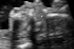
 |
| 3D-acquired fetal spine image, rendered view. To the left is the nonprocessed original, in the middle is the 3D image-enhanced spine, and to the right is the respective 2D-enhanced image. Note the clear delineation of the fetal spine in the 3D-enhanced image. Images courtesy of Dr. Anders Selbing, PhD.1 |
Most radiologists have been trained with 2D technology; combined with a lack of clinical evidence for 3D/4D in the past, this dynamic has led to clinician bias and low demand for technology improvements. With 3D acquisitions, however, the whole anatomy can be acquired in one single scan for most clinical applications. This may facilitate faster diagnosis and ultimately spark increased demand.
Benacerraf showed that a quick acquisition is sufficient to reconstruct the three orthogonal planes and show the cavity in each orientation desired.3 3D scans also provide access to the C plane, the plane perpendicular to the probe; this is not possible with 2D scanning techniques. For example, Tutschek described that "the vermis can be seen in the reconstructed plane in the sagittal view."2 Provided basic training on 3D scanning technique, less experienced radiographers can efficiently acquire accurate anatomical volumes, as stated by Selbing.1
Hardware updates
The graphics processing unit (GPU) is increasingly being used not only for gaming, but also for general purpose computing, either replacing the CPU or working with it. As GPU technology rapidly develops and standard programming languages emerge, it increases the potential to fully leverage 3D/4D acquisitions and 3D image enhancement software.
The rapid development of powerful and affordable hardware, combined with increasing demand from clinicians, will be a strong driver for OEMs to invest in powerful 3D technology. OEMs can easily upgrade to 3D/4D ultrasound probes to keep up with 3D/4D imaging software.
When combined with the appropriate hardware, today's software can cost-effectively deliver advanced capabilities that previously required expensive special-purpose hardware. For example, ultrasound scan conversion was originally a process completed by hardware. This can now be accomplished with advanced image processing software.
Obstetrics and gynecology are the strongest clinical applications for 3D/4D, with proven clinical benefits as described above. However, other clinical applications such as general radiology and urology are also increasingly in need of 3D acquisitions and are growing accordingly. As long as clinician demand remains on the rise, OEMs will have no choice but to invest in hardware capable of supporting 3D/4D imaging.
With years of accumulated clinical evidence and greater demand/advancements for hardware upgrades, 3D/4D will undoubtedly take major steps in 2013 to become the industry standard and support accurate, efficient, and real-time diagnoses.
References
- Selbing A. Interview on 3D obstetrics; October 2012.
- Tutschek B. 3D ultrasound approach to CNS evaluation. Presented at: International Society of Ultrasound in Obstetrics and Gynecology World Congress; September 10, 2012; Copenhagen, Denmark.
- Benacerraf B. 3D in gynecology. Presented at: International Society of Ultrasound in Obstetrics and Gynecology World Congress; September 11, 2012; Copenhagen, Denmark.
- Chiappa V. A possible role of 3D-ultrasound in the assessment of parametrial infiltration in cervical cancer. Presented at: International Society of Ultrasound in Obstetrics and Gynecology World Congress; September 2012; Copenhagen, Denmark.
- Goldberg B, Forsberg F, Lev-Toaff A. Image processing of 3D/4D ultrasound images; 2009.
As senior application manager at ContextVision, Isabelle Wegmann Hachette leads strategic product development and handles technical account management and installations for customers worldwide. She has a Master of Science degree in electrical engineering from the image processing laboratory at Linköping University, where she also served as university lecturer.
The comments and observations expressed herein do not necessarily reflect the opinions of AuntMinnie.com, nor should they be construed as an endorsement or admonishment of any particular vendor, analyst, industry consultant, or consulting group.