Flat colorectal lesions aren't easy to detect at virtual colonoscopy. But a group from Japan did quite well finding them after patients underwent bowel cleansing and automated insufflation before CT scanning, according to a new study in Acta Radiologica.
Using virtual colonoscopy (also known as CT colonography or CTC), the study team detected flat lesions 6 mm and larger with more than 87% sensitivity -- not much less than the 91.7% sensitivity clocked for the more common polypoid lesions in the same size range.
Sensitivity was far lower for flat lesions smaller than 5 mm, the authors cautioned; however, CTC studies almost always ignore findings smaller than 5 mm, owing to limitations in spatial resolution and the paucity of clinically relevant findings in diminutive polyps, which rarely harbor advanced histology. There were no significant differences in detection sensitivity between different colon regions.
The study was conducted by Dr. Takashi Sakamoto, Dr. Katsuhiko Mitsuzaki, Dr. Daisuke Utsunomiya, and colleagues from Saiseikai Kumamoto Hospital and Kumamoto University in Japan (Acta Radiol, July 20, 2012).
Overcoming challenge of flat polyps
Previous studies have boosted detection sensitivity for flat polyps by adding fecal tagging agents to the CTC protocol, the authors wrote.
"Although the detection of flat polyps by CTC is compromised by the lack of direct mucosal visualization, this disadvantage may be overcome by techniques such as bowel preparation and colonic distension," they wrote.
 |
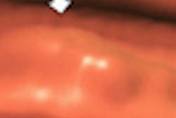
| Virtual endoscopic image at screening CTC shows a flat colon polyp, which was confirmed on optic colonoscopy. Image courtesy of Dr. Takashi Sakamoto. |
Previous studies of patients with colorectal cancer have reported flat lesions (defined for the current study as having a flat surface and a height not exceeding 3 mm) as relatively common, constituting about 10% of lesions in patients with advanced neoplasia, according to Sakamoto and colleagues.
Patients underwent a one-day bowel preparation on the day of the exam that included polyethylene glycol (1,600 mL) followed by sodium and meglumine amidotrizoate, a mixture of bowel cleansing agent and water-soluble iodinated contrast. Finally, an oral water-soluble contrast agent was administered for fluid tagging.
In all, 351 screening subjects (236 men, 115 women; average age, 51.4 ± 10.5 years) were included in the study, with optical colonoscopy performed before virtual colonoscopy on the same day. Before CT scanning, an automatic insufflator was used to administer CO2 for bowel distention (average volume for optimal distention, 1,958 ± 983 mL). After administration of an antispasmodic agent, a scout radiograph was obtained to determine sufficient colonic distention.
Prone and supine CTC images were acquired on a 64-detector-row scanner (Aquilion 64, Toshiba Medical Systems) at 120 kV and 100 mA for a total mean dose of 5.7 mGy per position. The images were reconstructed at a 0.5-mm slice thickness and examined on a workstation (Ziostation N610 1.21b, Amin) using multiplanar reformatting, volume rendering, dissected colon, and virtual endoscopic images. Two experienced readers, a board-certified radiologist and a gastroenterologist qualified as a supervising physician, examined the studies blinded to clinical data, interpreting the images in consensus.
The readers recorded the region, size, and morphology of each lesion. CTC findings 2 mm and larger were compared with optical colonoscopy as the gold standard, the study team wrote.
Virtual colonoscopy detected flat lesions 6 mm and larger with 87.5% sensitivity versus 91.7% for polypoid lesions in the same size range. VC's performance was lower for smaller lesions, with an overall detection sensitivity of 47.6% for flat lesions 2 mm and larger; this was significantly lower than VC's overall sensitivity for polypoid lesions (64.1%, p < 0.01). In all, 22 flat lesions and 150 polypoid lesions were missed at CTC.
CTC detection sensitivity for flat vs. polypoid lesions
|
The detection of flat polyps is important with virtual colonoscopy screening because small flat lesions are more likely to be a more advanced stage than polypoid lesions, the authors wrote when discussing their results. Sensitivity and specificity were relatively high, and there was no significant difference between different regions of the colon, they added.
CTC conditions were optimized by fecal tagging, and also by the suction removal of residual fluid during colonoscopy, which preceded CTC in every patient. Another unique aspect of the study was its evaluation of polyps smaller than 5 mm, performed because "their natural history remains to be elucidated," Sakamoto and colleagues wrote.
"Due to the thin-slice collimation of 64-row multidetector CT systems and the establishment of optimal conditions by careful bowel preparation, 56.6% of all lesions ≤ 5 mm were detected," the authors wrote. "Consequently, we consider the sensitivity of CTC for such small lesions acceptable. Our results document that 91.3% of polyps measuring ≥ 6 mm, the targets for treatment and detailed examination, can be detected at CTC screening."