Today, diagnostic radiology and radiation oncology are distinctly separate disciplines, each with their own specialized practitioners, professional societies, academic journals, and congresses. But things weren't always that way -- in fact, the early pioneers of radiology began experimenting with the new rays for therapeutic purposes almost as soon as they were discovered.
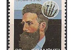
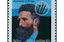
When Wilhelm Conrad Roentgen wrote his article about discovering invisible x-ray images on glass and fluoroscopic devices in November 1895, the scientists and doctors who followed his examples had the same ambitions for views of bones and segments of human anatomies. The use of vacuum tubes in the early years required as long as an hour to record the x-ray images of any body part.
Within the first few years, reports in medical journals commented that exposure to x-ray tubes caused changes to the exposed elements of humans. Generally, the extensive x-ray beam caused two kinds of effects to patients who received doses. One was that the x-ray beam seemed to cause correction of skin defects, muscular injuries, lupus, arthritis, and even the presence of superficial cancer growths.
However, the other was that extended exposure to x-rays was seen to cause skin rashes, and to cause the beginning of cancer growths. Some of the doctors and nurses who conducted frequent x-ray studies also noticed that they began to have external cancer growths. In the first decade, the general Roentgen gadgets made images. But the vacuum tubes also sprayed x-ray beams around the whole room where examinations were performed.
One of the other dilemmas in controlling early x-ray beams was a lack of any scientific measurement to detect the volume of x-ray exposure. And, in that same early decade, there were no definitive conclusions about the proper volume of radiation exposure to limit injuries or to retard abusive doses.
It was in 1913 that William Coolidge, an engineer at the General Electric Company, devised a more sophisticated x-ray device in which the vacuum tube was housed in a metal shielding container. An adjustable aperture allowed the operator to focus the beam on a chosen part of a patient's anatomy and the x-ray machine was left active for several more minutes to focus on exposed cancers.
In the years after acceptance of the basic Coolidge tube, the scientist was back in his laboratory working on more powerful x-ray beams to have a stronger effect on cancer treatment, particularly for many tumors that were located deeper inside a patient.
The radium approach
Just a year after Roentgen's discovery of x-rays in 1895, a French physicist, Henri Becquerel, discovered that pitchblende, a natural ore containing radioactive substances, might also provide refined materials that could be extracted and applied to human beings.
His close colleagues in Paris, Pierre and Marie Curie, worked to extract the radioactive energy from pitchblende. A year after Roentgen received the first Nobel Prize, the Curies also received a Nobel Prize for the development of radium. Tiny amounts of radium could be extracted in a complex and expensive process. And radon, the radioactive gas which leaked from radium, could be captured, placed in metal or glass tubes, and used to treat cancer patients.
In the early 1900s, the son of James Douglas died of cancer. James Douglas lived in New York City, where he was the owner of a large mining company with business in many parts of the U.S. He was a trustee of the Memorial Cancer Hospital and also Cornell Medical School.
With an ambition to find a better way to produce radium, he directed his mining company to excavate carnotite ore from Colorado and extract radium. By 1912, he had contributed two grams of radium to Memorial Hospital, and in 1916, another two grams; shortly, the amount became some 80% of radium available in the U.S. The hospital had more radium than anyone else in the country, and it applied it to the treatment of cancer patients.
Powerful cancer treatment
Even while William Coolidge was building x-ray devices operating at 200 kV to 400 kV, the concept of radiologists specializing in cancer treatment led to the conclusion that radiation tools which could generate beams of 1 million kV could impact cancers in any internal part of a patient's body.
One of the most potent radiation sources in the 1930s was the electron-beam therapy accelerator devices contrived by Robert Van de Graaff at the Massachusetts Institute of Technology in Boston. Several other x-ray equipment companies had experimented with the million volt generators, and some had produced medical betatrons.
In the late 1920s, Ernest Lawrence, a senior physicist at the University of California, Berkeley began building the first artificial cyclotrons in the U.S. and began cooperating with Robert Stone, the chief of radiology at the University of California Medical School, across the bay in San Francisco.
One of Lawrence's team members built a 1-million-volt therapy device for Stone to use in the 1930s. As the cyclotrons became more potent, they were able to generate artificial radioisotopes, notably sodium-24 and phosphorus-32. When the cyclotrons generated neutrons, Stone took cancer patients across to the university campus in Berkeley to treat them on a cyclotron. The cyclotrons did not prove to be as effective as some of the other high-energy x-ray devices.
Before World War II, the efforts to develop artificial radioisotopes were too costly for general acceptance. However, during the war, the U.S. government had created a secret government agency to build atomic bombs. And at the end of the war, the federal government legislated the Atomic Energy Commission to create and share artificial isotopes.
Bob Stone had been a consultant to the atomic bomb project and he returned to the University of California at the end of the war. In 1950, he obtained funds from the Atomic Energy Commission to build a 70-million-volt synchrotron to treat cancer patients at his facility in San Francisco. That device was not a crowning success.
Shortly after World War II, Henry Kaplan, a radiologist focusing on cancer treatment, became the chairman at Stanford University's medical school, which at the time was in San Francisco. He sought cooperation from physicists at Stanford for the creation of potent linear accelerators. The first one was installed in his department.
In the late 1950s and early 1960s, researchers developed a new cancer treatment device using a pellet of cobalt-60 on a rotating device at the MD Anderson Cancer Hospital in Houston, and at the medical school at the University of Saskatchewan in Canada.
Linear accelerators with an energy of several million volts have become the most common radiation treatment sources in the past decades. The equipment to direct powerful x-ray beams to cancer patients is now managed by sophisticated treatment planning surveys and protocols. In half a century, medical radiation has separated between the diagnostic and interventional tools and techniques and the distinct creation of the discipline of radiation oncology.
Otha W. Linton, MSJ, retired in 1997 as the associate executive director of the American College of Radiology (ACR) after 35 years. He also served as executive director of Radiology Centennial in 1995. Mr. Linton holds a bachelor's degree in journalism from the University of Missouri and a Master of Science in journalism from the University of Wisconsin. His work has been published widely in the U.S. and abroad, and he is a regular contributor to several journals including Academic Radiology, the American Journal of Roentgenology, Radiology, and the Journal of the American College of Radiology. He joined the ACR staff in 1961 and had a key role in its growth. Over the years, his responsibilities with the ACR included government affairs, public relations, marketing, publishing, industrial liaison, and international relations. Just before his ACR retirement, he became the executive director of the International Society of Radiology and continues in that role. Also, since his retirement, he has written and published 14 histories of radiology societies and academic centers.