Training nonmedical personnel to perform simple volume ultrasound scan protocols can produce effective results, according to a research team led by Dr. Brian Garra of the University of Vermont School of Medicine in Burlington.
"Protocols for use by nonmedical personnel are feasible for screening," Garra said.
Garra presented the group's findings at the 2008 American Institute of Ultrasound in Medicine (AIUM) meeting in March.
While ready access to medical imaging has revolutionized diagnosis in the U.S. and other technologically advanced countries, the same can't be said for developing countries, Garra said. Rural inhabitants of those nations frequently have little or no access to advanced medical imaging technologies.
To leverage the low cost and power requirements of ultrasound to bring imaging to rural villages in these countries, the research group has developed a plan to donate or provide extremely low-cost ultrasound systems to a number of villages and/or small clinics. Power and an Internet uplink will be provided, and a local nonimaging person will be trained to perform screening volume scans.
"If it was an imaging person, that would be great," he said. "But we're not dependent on that."
This person will perform screening exams and upload them to an interpretation server via a cellular or satellite link to the Internet. A volunteer sonologist will then interpret the studies, and a translated report along with recommendations will be returned to the village.
The working group involved in this project includes the University of Vermont/Fletcher Allen Health Care (FAHC), GE Healthcare, Philips Healthcare, Vivalog Technologies, Princeton University, and McKesson.
The scan protocols that have been designed are volume imaging-dependent, and they involve four to eight imaging sweeps performed using surface anatomy as a reference, according to Garra.
"So there's no knowledge of internal anatomy required," he said.
Scanning presets are critical; the scanner has to automate most machine adjustments that the sonographer or physician would ordinarily make, Garra said. Image labeling has to be automated as well.
Four protocols were developed to scan the thyroid, gallbladder, kidney, and female transabdominal pelvis. To test the protocols, pairs of volunteers without imaging experience underwent a 20- to 30-minute training session for each protocol.
Volume sweeps were conducted using a Logiq i scanner (GE, Chalfont St. Giles, U.K.) and sent to a PACS for evaluation by two physicians and one sonographer. The evaluation criteria included a percentage visualization of each organ in the area of the examination, the quality of visualization on a six-point scale (0 = no visualization), and a quality score for the study based on an AIUM accreditation checklist. A 70% quality score was considered passing.
The protocols were tested on 20 volunteer subjects with no known pathology. Some volunteers agreed to scan the same subject 10 times to assess reproducibility, and several subjects agreed to be scanned by up to 10 individuals to assess interexaminer variability.
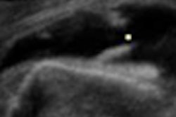
In the thyroid, the right lobe had an average of 89.3% anatomic visualization as judged by the panel and a visualization quality of 3.1 (out of 6). The left lobe had an 89% anatomic visualization, with a quality score of 3.1, while the isthmus had a 94.1% anatomic visualization and a quality score of 3.5. The adjacent soft tissue had a 90.1% anatomic visualization and a quality score of 3.2.
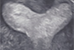
As for the gallbladder, the average anatomic visualization percentage was 74.3%, with a quality score of 2.69. The right liver had an average visualization percentage of 55.2% and a quality score of 2.91, while the hepatic veins had a visualization percentage of 68% and a quality score of 2.73. The portal vein had a visualization percentage of 74.2%, with a quality score of 2.76.
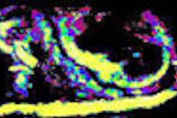
In the kidney protocol, findings included a 62.8% visualization percentage for the longitudinal right view and a quality score of 2.11. The transverse right view had a visualization percentage of 59.8% and a quality score of 2.29.
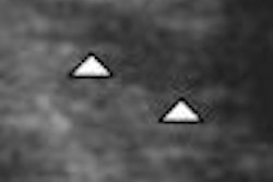
The fourth protocol, the female pelvis, yielded results such as a 59.3% anatomic visualization percentage and a quality score of 1.86. The endometrium had a 33.7% anatomic visualization and an average quality score of 1.79.
Dr. Garra noted that his visualization percentage evaluations and quality scores were higher than the panel's.
The researchers have also begun a study to evaluate the repeatability of the scanning protocols. While limited data is available so far, the researchers have found a 5% to 20% variation of the panel scores from exam to exam, Garra said.
"I think that's pretty reasonable," he said.
The group found that the pelvis scans had a big variation, which might have been caused by some patients having full bladders, according to Garra.
In this program, training the trainers is most important because the precise setup and transducer positioning are critical when the person doing the scanning doesn't know where the organ is, he said.
Quality improvement will also be an ongoing issue in this program, Garra said. "We're going to have to have quality monitoring on an ongoing basis ... so that people don't forget how to do these protocols correctly."
By Erik L. Ridley
AuntMinnie.com staff writer
April 29, 2008
Related Reading
3D ultrasound can improve life for sonographers, save money, March 18, 2008
Job survey shows growing sonographer demand, February 28, 2008
Keeping sonographers by improving working conditions, July 5, 2007
Sonographers: Highly experienced, vastly underappreciated, February 6, 2007
Head and neck surgeons can handle ultrasound, journal article says, January 5, 2007
Copyright © 2008 AuntMinnie.com