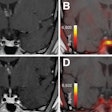
A calcium-sensitive contrast agent, married with superparamagnetic iron oxide particles (SPIO), could make MR studies of the brain on a molecular level feasible, according to researchers from the Massachusetts Institute of Technology in Cambridge.
Functional MRI (fMRI) techniques can capture blood flow in the brain, but these hemodynamic changes occur seconds after the neuron has fired. As a result, current techniques are not equipped to measure precise neuronal activity. Optical imaging, which is sophisticated enough to capture this activity, still suffers from a restricted field-of-view.
"MRI studies of calcium dynamics could, in principle, complement optical approaches by offering both greatly expanded coverage and depth penetration in vivo," wrote Tatjana Atanasijevic, a graduate student in the departments of nuclear science and engineering at MIT. Her co-authors included Alan Jasanoff, Ph.D., an associate member of the McGovern Institute for Brain Research at MIT (Proceedings of the National Academy of Sciences, October 3, 2006, Vol. 103:50, 1407-1412).
For this study, the group used a 4.7-tesla research scanner (Avance, Bruker BioSpin, Billerica, MA) and SPIO nanoparticles coated with a streptavidin protein. To construct the MR calcium sensors, two molecular binding agents, biotin-CaM and biotin-M13, were produced and attached to the SPIOs.
The imaging protocol for the mica samples consisted of a spin-echo pulse sequence with multiecho acquisition to collect images at several parallel echo times, the authors explained.
According to the results, "the images indicate that the MRI signal ... is significantly greater when calcium is present, and that the effect is mot pronounced at longer echo time while still maintaining excellent signal-to-noise ratio," they stated. "MRI measurements collectively show that calcium-induced clustering of our sensors corresponds to decreases in T2 relaxivity (producing greater T2-weighted MRI signal), in contrast to results obtained with smaller mononuclear iron oxides."
Jasanoff's group is currently working on noninvasive ways to deliver this calcium sensor to brain cells in vivo in small animal studies.
By Shalmali Pal
AuntMinnie.com staff writer
December 6, 2006
Related Reading
MRI offers in-depth, biodynamic view of cellular therapy, November 2, 2005
A new use for SPECT/CT: Tracking stem cell migration, October 7, 2005
Copyright © 2006 AuntMinnie.com


