SAN DIEGO - A fused cardiac image rendered through the combination of dual-head SPECT with 64-slice CT technology by researchers from the University Hospital Zurich in Switzerland was awarded the Society of Nuclear Medicine's (SNM) 2006 Image of the Year on Monday afternoon.
The image, part of the study "Comparison of 64-slice spiral CT angiography and myocardial perfusion imaging in noninvasive evaluation of functionally relevant coronary stenosis," was selected by Dr. Henry Wagner, SNM past president, historian, and professor of environmental health sciences at Johns Hopkins University in Baltimore.
The image, which shows both the coronary arteries and blood flow to the heart, allows for the identification of heart disease and shows the complementary nature of the two imaging modalities, Wagner said. The study shows that purely anatomical imaging of the heart is often insufficient to identify coronary lesions, underlining the need for a combined assessment that may be made possible with hybrid fusion imaging such as SPECT/CT or PET/CT.
The study from which the Image of the Year was selected prospectively compared the accuracy of 64-slice CT (LightSpeed VCT, GE Healthcare, Chalfont St. Giles, U.K.) angiography (CTA) with myocardial perfusion imaging (MPI) using Tc-99m tetrofosmin-SPECT (Millennium VG Series Hawkeye, GE Healthcare) as the gold standard for detecting functionally relevant coronary lesions.
 |
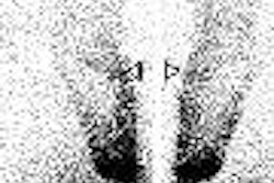
| The upper row shows two short-axis slices after pharmacological stress; the lower row shows the same slices when the body is at rest in conventional cardiac SPECT with Tc-99m tetrofosmin as a radiotracer. Arrows indicate a small perfusion defect on the back side of the heart -- visible only on the stress images -- showing ischemia in this region of the heart wall. Image courtesy of Drs. Oliver Gaemperli and Philipp Kaufmann, University Hospital Zurich, Switzerland. |
The group examined 100 consecutive patients with suspected or known coronary artery disease, and performed electrocardiographic (ECG)-gated MPI and 64-slice CTA on each patient.
The SPECT protocol, according to Dr. Oliver Gaemperli of the research team, was to image at pharmacologic stress (300 MBq of adenosine) and rest (900 MBq of Tc-99m tetrofosmin). The group used a matrix of 64 x 64, low-energy collimators, a 20% window, a step-and-shoot acquisition at 180° with 3° steps, a 20-second dwell time on a dual-head camera with 90° between the heads, and ECG-gated acquisition with a 50% window of acceptance.
The 64-slice CTA protocol employed collimation of 64 x 0.625 mm, a z-axis coverage of 40 mm, a gantry rotation time of 0.35 seconds, 80-120 mL of iodine-based contrast media delivered via dual-component power injector with a 50 mL saline chaser bolus, 120 kVp, and the tube current was ECG-modulated between 280 mAs and 750 mAs. Reconstruction was conducted in 0.625-mm increments, while the ECG-gated reconstruction was at 5% to 95%, Gaemperli said.
 |
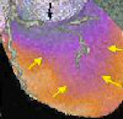
| The Society of Nuclear Medicine's 2006 Image of the Year is "a beautiful fusion image of a 64-slice CT with SPECT," noted Dr. Henry Wagner, who selected the Zurich University study that was given the award. The images show a SPECT/CT fusion of the anterior and posterior heart and its coronary arteries obtained by 64-slice CTA with the functional information about the corresponding perfusion from cardiac SPECT (represented by the orange and yellow colors) superimposed. Images courtesy of Drs. Oliver Gaemperli and Philipp Kaufmann, University Hospital Zurich, Switzerland. |
The images from the two modalities were fused utilizing a CardIQ Fusion software package (GE Healthcare). The resulting renderings accurately pinpoint a blood-flow defect to its corresponding artery, according to the research team.
The group said it found CTA to be an excellent tool to rule out relevant coronary artery disease, but noted that not every plaque or lesion in the coronary arteries causes significant reduction of blood flow to the heart. However, MPI with SPECT does visualize blood flow to the heart.
The study results underline the importance of a combined assessment with CTA and SPECT to identify those lesions that may need urgent revascularization and those that may be treated conservatively, the researchers said.
By Jonathan S. Batchelor
AuntMinnie.com staff writer
June 6, 2006
Related Reading
Dr. Henry N. Wagner Jr.: A history in nuclear medicine, June 4, 2006
3D rendering captures SNM's Image of the Year, June 21, 2005
Brain image database wins Image of the Year, June 22, 2004
LSO PET/CT study wins Image of the Year, June 24, 2003
PET, SPECT studies chosen as SNM images of the year, June 18, 2002
Copyright © 2006 AuntMinnie.com