PET imaging has shown a link between serotonin brain activity in early Alzheimer's disease patients to beta-amyloid plaque deposits, a hallmark of the condition, according to a study published online on January 6 in NeuroImage: Clinical.
Researchers at Johns Hopkins University in Baltimore compared brain PET images of a serotonin transporter called 5-HTT and amyloid plaque in a group of patients and healthy controls. They found a pattern of decreased 5-HTT and increased beta-amyloid deposits separated people with mild cognitive impairment (MCI) from controls.
The finding could open avenues for new treatment approaches, the authors wrote.
"Understanding the role of the serotonin system in the preclinical course of [Alzheimer's disease] and the relationship to beta-amyloid deposition may have therapeutic implications," wrote a team led by Gwenn Smith, PhD, co-director of the university's Center for Translational Molecular Imaging.
Serotonin plays a significant role in normal brain function and modulates fundamental behaviors, including mood, sleep, appetite, and cognition. Degeneration of the serotonin system has been observed in previous studies in Alzheimer's and patients with MCI.
Animal studies have shown serotonin degeneration before the development of widespread beta-amyloid deposits. For the study, Smith and colleagues sought to confirm this same association in human participants, comparing imaging results in 45 patients with MCI and 35 healthy older adults who completed clinical and cognitive evaluations and 5-HTT and amyloid PET scans using established radiotracers.
According to the findings, a pattern of lower cortical, subcortical and limbic 5-HTT availability and higher cortical beta-amyloid deposits on the PET scans distinguished the MCI participants from the healthy older controls, a statistically significant correlation (p < 0.00001).
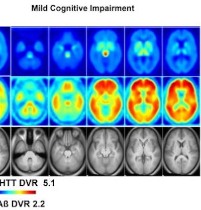
 PET scans of 5-HTT and beta amyloid in healthy controls and individuals with MCI. Mean distribution volume ratio images of healthy controls (left panels) and MCI (right panels) and an MRI template (bottom panel). Image courtesy of NeuroImage: Clinical through CC BY 4.0.
In addition, the spatial covariance pattern of the two markers corresponded with neuropsychiatric symptoms in the MCI group, the researchers reported.
"Greater expression of this pattern was correlated with greater deficits in memory and executive function in the MCI group, [but] not in the control group," the researchers wrote.
The researchers noted that the study's goal was not to reveal a new biomarker for early Alzheimer's but to open avenues for further research to explain the association between serotonin degeneration and beta-amyloid deposits.
"An understanding of the role of serotonin degeneration in relation to [Alzheimer's disease] pathology in the preclinical stages of [Alzheimer's disease] is critical," Smith and colleagues concluded.