PET/MRI imaging may be a valuable option over PET/CT for planning surgery in women with endometrial cancer, according to a study published September 1 in BMC Cancer.
A Chinese team in Shenyang compared how well the methods performed at identifying the severity of tumors prior to surgery in a group of patients with endometrial cancer. PET/MRI was more accurate than PET/CT at classifying early tumors, as well as visualizing cancer invasion in surrounding tissue, the group found.
"Our findings may help physicians in selecting more appropriate individualized treatment plans for patients," wrote corresponding author Dr. Hongzan Sun, PhD, a radiologist at Shengjing Hospital of China Medical University, and colleagues.
Endometrial cancer is typically diagnosed by ultrasound and visual exams of the endometrium with a hysteroscope, followed by a biopsy. After a woman is diagnosed, the next step is staging, wherein imaging is performed to determine how advanced the cancer is and how best to treat it.
MRI and PET/CT are two standard imaging techniques for staging endometrial carcinoma patients. Each has their advantages. MRI may be better at identifying primary tumors, while PET/CT identifies more metastatic disease, according to the authors.
Hybrid PET/MRI imaging is a relatively new approach that combines the anatomical and functional advantages of both techniques in a single scanner. In this study, the authors explored the value of PET/MRI for the first time in endometrial carcinoma staging and compared it to PET/CT.
The researchers culled data from 57 women with biopsy-proven endometrial carcinoma who underwent both F-18 FDG PET/CT and F-18 FDG PET/MRI (Signa PET/MRI, GE Healthcare) followed by surgery from December 2017 to January 2021 at Shengjing Hospital. The patients' tumors were classified as stage I-IV according to a standard system established by the International Federation of Gynecology and Obstetrics (FIGO).
The diagnostic performance of PET/MRI and PET/CT was compared by three physicians, each with double board certifications in radiology and nuclear medicine and more than five years of experience.
The overall accuracy of FIGO staging was 86% (49/57) for PET/MRI and 77.2% (44/47) for PET/CT, according to the findings. PET/MRI overstaged the actual FIGO stage in five patients, whereas PET/CT overstaged the actual FIGO stage in nine patients.
"Our results revealed that PET/MRI staging, which was performed using an integrated PET/MRI scanner, had an excellent diagnostic performance for endometrial carcinoma," the authors wrote.
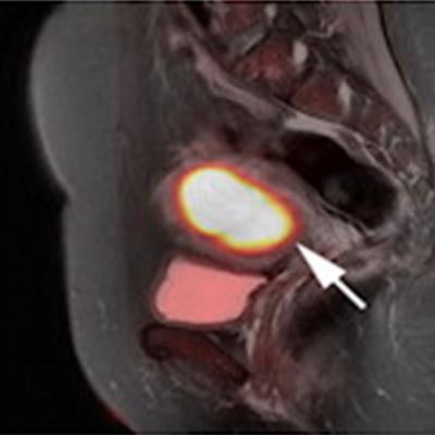
 FIGO stage II pathologically confirmed moderately differentiated adenocarcinoma in a 57-year-old female patient. (A) Axial PET/MR images. (B) Axial MR T2-weighted images. (C) Axial PET/CT images. (D) Axial CT images. (E) Sagittal PET/MR images. (F) Sagittal PET/CT images. PET/MRI and MRI show the endometrial carcinoma invading the cervical stroma. PET/CT also shows the endometrial carcinoma invading the cervical stroma, yet the extension of the tumor is difficult to determine. The PET/MRI and PET/CT staging were consistent with the pathological stage, namely FIGO stage II. The white arrow indicates endometrial carcinoma. Image courtesy of BMC Cancer.
In addition, an analysis of quantitative measures based on F-18 FDG PET/MRI radiotracer uptake by the tumors and nearby tissue were significantly different between stage I and stage III tumors, the authors wrote.
"PET/MRI had higher accuracy for endometrial carcinoma staging, mainly for FIGO stage I tumors. This was mainly owing to PET/MRI having higher diagnostic accuracy for detecting the depth of myometrial invasion," they wrote.
Ultimately, while pathological examination is the gold standard for endometrial carcinoma staging, biopsies can be traumatic to patients, and diagnosing the depth of tumor invasion in adjacent tissue and organs is difficult, the researchers wrote.
Thus, precise staging based on imaging has received greater research attention in recent years, with hybrid PET/MRI leading the way in studies of clinical gynecological malignancies, they wrote.
"PET/MRI may be a more valuable diagnostic tool to replace traditional imaging modalities for preoperative staging in patients with endometrial carcinoma," Sun and colleagues concluded.