Biopsy guided by contrast-enhanced ultrasound (CEUS) is effective, safe, and more accurate than conventional ultrasound-guided biopsy when it comes to diagnosing focal liver lesions, a multicenter study published August 2 in Radiology found.
A team led by Dr. Wei Wu from Peking University Cancer Hospital and Institute in Beijing wrote that this finding especially goes for lesions smaller than 2 cm and for hepatocellular carcinoma diagnosis. Contrast-enhanced ultrasound-guided biopsy can be considered an option for patients.
"Given the high sensitivity of CEUS-guided biopsy in detection of malignancies, clinical follow-up of lesions diagnosed as benign at biopsy can be considered an accurate approach," wrote Wu and co-authors.
Percutaneous biopsy is often needed to confirm diagnosis of focal liver lesions, with ultrasound typically used to guide biopsies and establish a histopathologic diagnosis of liver tumors. The researchers noted that about 10% of results when using this method are either uncertain or reported as false negative.
CEUS has been explored in recent years in improving the diagnostic accuracy of percutaneous biopsy. However, data have largely come from small samples, including retrospective and single-center studies.
Wu et al wanted to compare the diagnostic performance of CEUS-guided biopsy with conventional ultrasound-guided biopsy for focal liver lesions in a prospective multicenter study.
They collected data between 2016 and 2019 from 2,056 study participants (1,297 men, 759 women) with an average age of 58 years. Focal liver lesions were detected with ultrasound, CT, or MRI and patients were planned for percutaneous biopsy. Patients were randomly assigned to undergo either conventional ultrasound- or CEUS-guided biopsy. From the total, 1,030 patients underwent biopsy in the conventional arm while 1,026 were assigned to the CEUS arm.
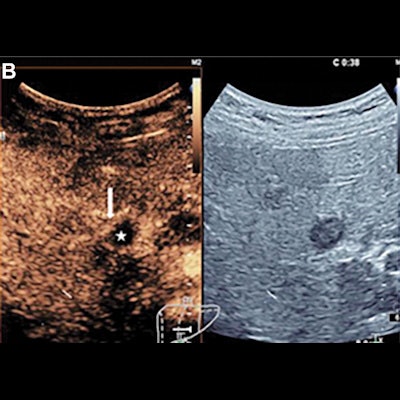
 CEUS images show a change of the biopsy target area in a 54-year-old woman who presented with a two-week history of epigastric discomfort. A pancreatic mass and focal liver lesion were detected with standard ultrasound. (A) Ultrasound shows a hypoechogenic nodule with a 1.6-cm diameter in the left medial lobe. The center of the lesion was chosen as the biopsy target area at ultrasound. (B) CEUS scans show ringlike enhancement (arrow) of the focal liver lesion in the arterial phase. There was no enhancement in the center of the lesion (★), which indicated necrosis. (C) Biopsy was performed in the ringlike enhancement area with CEUS guidance in the arterial phase. (D) Pathologic analysis allowed for confirmation of an adenocarcinoma (Hematoxylin-eosin stain; original magnification, ×100). Image courtesy of the RSNA.
| CEUS-guided biopsy vs. conventional ultrasound-guided biopsy for focal liver lesions | |||
| Conventional arm | CEUS arm | p-values | |
| Overall accuracy | 93% | 96% | 0.002 |
| Correct identification | 92% | 96% | 0.002 |
| Negative predictive value | 57% | 74% | 0.001 |
| Accuracy (lesions smaller than 2 cm) | 88% | 96% | 0.004 |
| Sensitivity (lesions smaller than 2 cm) | 87% | 95% | 0.007 |
The researchers also found that the accuracy of CEUS-guided biopsy (93%) was superior to that of standard ultrasound-guided biopsy (80%) among lesions smaller than 2 cm (p = 0.008). However, the two methods were comparable when it came to liver metastases (p = 0.63).
The study authors noted that the target lesions or areas were changed after CEUS in 257 cases, which they noted "likely" resulted in a higher biopsy accuracy in the CEUS-guided biopsy group. They added that CEUS can be used to identify vascularized areas of a lesion and that hypoenhancement in the late phase is a well-known CEUS pattern of malignancy.
"Thus, our data enabled us to confirm that CEUS is very helpful in the identification of accurate puncture areas and in guiding the biopsy," the team wrote. "CEUS-guided biopsy may be particularly useful in focal liver lesions with suspected necrosis."
The researchers also noted that the decision on further patient care should not be based on the guided biopsy results alone and should account for disease history and imaging characteristics.