Deep-learning analysis of joint MRI exams is an up-and-coming application of artificial intelligence (AI) that could dramatically change the practice of musculoskeletal radiology, according to a review article published September 1 in Skeletal Radiology.
The technology shows potential for supporting radiologists in their musculoskeletal MRI interpretations, wrote Dr. Benjamin Fritz of the University of Zürich in Switzerland and colleague Dr. Jan Fritz of New York University.
"Contingent upon future studies showing the clinical utility of deep-learning algorithms, artificial intelligence may eventually translate into clinical practice to assist detection and characterization of various conditions on musculoskeletal MRI exams," the two wrote.
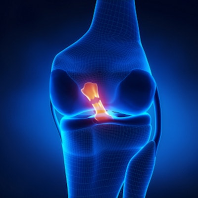
Existing AI algorithms for musculoskeletal imaging assess cruciate ligament tears, meniscus tears, and rotator cuff injuries on MRI exams of the knee and shoulder, but human readers still perform better, the authors noted.
Is there room for improvement of AI algorithms in musculoskeletal MRI? Yes, Fritz and Fritz wrote.
"Deep learning-based segmentation algorithms of nerves, bones, and muscles are promising tools for automated quantification and potentially improved reproducibility and efficiency," they wrote.
The two explored the current state of deep learning for musculoskeletal MRI exams through an assessment of clinical literature. Among other studies, their review included nine papers on AI for anterior cruciate ligament tear detection and six papers on AI for automated meniscus tear detection.
They also evaluated research on the use of deep learning with "other internal derangement abnormalities" such as osteoarthritis, joint effusions, iliotibial band syndromes, bone contusions and fractures, and posterior cruciate ligament tears; shoulder MRI applications (rotator cuff, long head biceps tendon, and articular cartilage injuries); ankle applications; MR neurography for imaging the sciatic nerve; and for identifying and categorizing musculoskeletal tumors.
Particularly for using AI to detect anterior cruciate ligament tears, Fritz and Fritz noted a specificity range of 89% to 100% for AI and a range of 86% to 98% for human readers. Sensitivity had similarly comparable ranges.
| AI versus radiologist readers for musculoskeletal MRI | ||
| Measure | Human readers | AI algorithm |
| Anterior cruciate ligament tear detection | ||
| Sensitivity | 91%-98% | 76%-100% |
| Specificity | 86%-100% | 89%-100% |
| Automated meniscus tear detection | ||
| Sensitivity | 82%-94% | 71%-91% |
| Specificity | 88%-90% | 74%-90% |
The bottom line? Deep learning for musculoskeletal MRI is an exciting frontier, according to Fritz and Fritz, who suggest that "at some point in the future, there may be deep-learning algorithms for MRI diagnosis of many different anatomical structures, injury patterns, and pathological conditions fully integrated into departmental workflows."
In fact, the two predict that, over the next decade, radiologists may shift from being the primary identifiers of abnormalities to "supervision and quality control of deep learning-based detection."
"Although musculoskeletal MRI interpretations of subspecialized radiologists have high accuracies, many years of training are usually required to attain proficiency for the broad range of musculoskeletal MRI exams," they concluded. "Disease-detecting deep learning algorithms may aid in providing expert-level interpretations for readers with less expertise and may also play a role in teaching residents and fellows."
Disclosure: Dr. Jan Fritz has received research support from Siemens, BTG International, Zimmer Biomed, DePuy Synthes, QED, and SyntheticMR. He is a scientific advisor for Siemens, SyntheticMR, GE Healthcare, QED, BTG, ImageBiopsy Lab, Boston Scientific, and Mirata Pharma and shares patents with Siemens Healthineers and Johns Hopkins University.