Scurvy -- an old but not forgotten disease -- was diagnosed on x-ray recently in a 2-year-old girl in Buffalo, NY, according to a report August 18 in the Journal of Imaging in Emergency Medicine.
Scurvy is caused by vitamin C deficiency, which must persist for eight to 12 weeks before symptoms develop. The disease has great historical significance, but it is almost unheard of in patients without a significant underlying predisposing condition that leads to dietary restriction, such as developmental delay or medically restrictive diets.
In this case report, the girl presented with a new petechial rash. On arrival, she was described as underweight and disheveled, and she could not bear weight due to bilateral leg pain. She had been experiencing intermittent fevers and joint pain for a month.
An initial oropharyngeal exam showed mucous membranes with bleeding and receding gingiva, while a dermatologic exam revealed scant scattered ecchymosis and bilateral petechiae with perifollicular hemorrhages most pronounced on her lower extremities, wrote the authors of the report.
In the girl's case, there was no prior diagnosis of developmental delay, they noted. Due to concern for neglect, they ordered a skeletal survey.
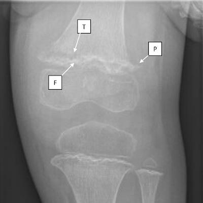
Subsequent images of the girl's distal femur and proximal tibia showed three common radiological findings in scurvy:
- Frankel lines -- dense zones of provisional calcification
- Pelkan spurs -- bony spurring at the periphery of the zone of metaphyseal calcification
- Trümmerfeld zone -- lucent bands in the metaphysis below the Frankel line
"These radiographic findings, in conjunction with the clinical findings of mucosal bleeding, perifollicular petechiae, and arthralgias, suggested a diagnosis of scurvy," wrote first author Dr. Matthew Wiese, a pediatric emergency medicine fellow at Oishei Children's Hospital.
 Radiograph of the patient's left knee showing dense zones of provisional calcification (F: Fraenkel line) with underlying lucent metaphyseal bands (T: Trummerfeld zone). Also identified is spurring at the distal femur and proximal tibia (P: Pelkan spurs) and a displaced fracture of distal femoral metaphysis. Image courtesy of the Journal of Imaging in Emergency Medicine.
The clinical and radiographic characteristics of scurvy are well defined, yet notably, the clinicians identified the disease without a key piece of information -- the girl's vitamin C level.
"Due to a lab processing error, the patient's ascorbic acid level did not result," they wrote.
Thus, scurvy can be a clinical diagnosis with history and physical exam findings confirmed by radiographs showing Frankel lines or Trümmerfeld zones, the team stated.
As for the patient, she rapidly improved after receiving vitamin C supplements, which in itself ultimately confirmed the diagnosis, the authors concluded.