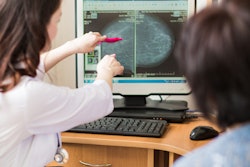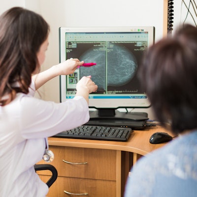
Returning to routine screening for BI-RADS 3 lesions on supplemental automated whole-breast ultrasound reduces the cancer recall rate "substantially," while also being unlikely to result in adverse outcomes, according to research published July 14 in the American Journal of Roentgenology.
Results from a prospective study led by Richard Barr, PhD, from Northeastern Ohio Medical University in Rootstown supports routine annual follow-up for BI-RADS 3 lesions at supplemental automated whole-breast ultrasound.
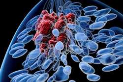
This method of ultrasound detects additional cancers in women with dense breasts, but it also identifies many BI-RADS 3 lesions that result in short-term follow-ups and biopsies, Barr and his team said. Researchers wanted to evaluate outcomes in patients recommended for return to routine screening for lesions assessed as BI-RADS 3.
The team looked at 2,257 women between 2013 and 2016 with an average age of 58 years old. Whole-breast ultrasound was scored as BI-RADS 1 in 1,186 of the women, BI-RADS 2 in 591, BI-RADS 3 in 395, and BI-RADS 0 in 85.
The study authors found that routine follow-up of BI-RADS 3 lesions detected on supplemental screening in women with dense breasts and any risk resulted in a recall rate of 3.8%, a biopsy rate of 0.5%, and a positive biopsy rate of 58.3%, with no missed cancers.
A total of 394 patients with BI-RADS 3 had a two-year follow-up, during which no cancer was diagnosed in the quadrant of the lesion.
The researchers also found that malignancy rates were not different between BI-RADS 1, 2, and 3, and the ultrasound recall rate was 3.8%. They also found that if short-term follow-up had been recommended for automated whole-breast ultrasound BI-RADS 3, the recall rate would have been 21.3%.
Barr and his team said that automated whole-breast ultrasound can help lessen variability by standardizing documentation, recording, and archiving of ultrasound images from the entire breast, except the axilla. They also said that images of similar quality can be obtained from adequately trained ultrasonographers and mammographers.